E-ISSN:2349-3267
P-ISSN:2349-5499

Case Report

Disorder

Pediatric Review - International Journal of Pediatric Research

2020 Volume 7 Number 3 March
Publisherwww.medresearch.in

Cornelia de Lange syndrome: A rare genetic disorder

Baburao Sonawane V.1*, Kotrashetti V.2, Bainade K.3, Vatkar A.4, Pansare A.5
DOI: https://doi.org/10.17511/ijpr.2020.i03.07

1* Vijay Baburao Sonawane, Associate Professor, Department of Pediatrics, D.Y. Patil University, School of Medicine, Nerul, Maharashtra, India.

2 V. Kotrashetti, Professor and Head of Unit, Department of Pediatrics, D.Y. Patil University, School of Medicine, Nerul, Maharashtra, India.

3 Kapil Bainade, Assistant Professor, Department of Pediatrics, D.Y. Patil University, School of Medicine, Nerul, Maharashtra, India.

4 Amit Vatkar, Assistant Professor, Department of Pediatrics, D.Y. Patil University, School of Medicine, Nerul, Maharashtra, India.

5 Abolee Pansare, Resident (PG), Department of Pediatrics, D.Y. Patil University, School of Medicine, Nerul, Maharashtra, India.

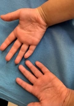
Cornelia de Lange Syndrome (CdLS) was first reported by Vrolik in 1849 and Brachmann in 1916, followed by Cornelia de Lange in 1933, after whom the syndrome is named. This disorder has a varied presentation but is mainly characterized by distinctive facial features, growth retardation, microcephaly, hirsutism, psychomotor delay, intellectual disability, and malformations of the upper limbs. Initial diagnosis is usually based on clinical features following specific diagnostic scoring systems. The precise prevalence of the disease is unknown but is estimated to be 1–10:100,000. Depending on the mutated gene, Cornelia de Lange syndrome (CdLS) can be inherited in an autosomal dominant manner, when it is caused by variations in the NIPBL, SMC2, or RAD21 genes, or it can have an X-linked inheritance when it is caused by variations in the SMC1A or HDAC8 genes. However, most cases (more than 99%) result from new (de novo) mutations, which means that are not inherited from the parents and occur in people with no family history of the conditionabout 30% of the people affected by the syndrome do not have any known cause. Many studies focused on the importance of neurologic findings and reported an incidence of epilepsy in CdLS ranging from 14% to 25%, especially in the classic and more severe form of the syndrome, but there is no data about its electroclinical features and long-term outcome. Life expectancy is relatively normal for people with CdLS and most affected children live well into adulthood. However, certain features of this condition, particularly severe malformations of the heart or throat, may decrease life expectancy in some affected people. The diagnosis is suspected clinically and later confirmed by clinical exome sequencing.

Keywords: Cornelia de Lange Syndrome (CdLS), Epilepsy, Developmental Delay, Clinical Exome Sequencing

Corresponding Author How to Cite this Article To Browse
Vijay Baburao Sonawane, Associate Professor, Department of Pediatrics, D.Y. Patil University, School of Medicine, Nerul, Maharashtra, India.
Email:
Sonawane VB, Kotrashetti V, Bainade K, Vatkar A, Pansare A. Cornelia de Lange syndrome: A rare genetic disorder. Pediatric Rev Int J Pediatr Res. 2020;7(3):152-156.
Available From
https://pediatrics.medresearch.in/index.php/ijpr/article/view/569

Manuscript Received Review Round 1 Review Round 2 Review Round 3 Accepted
2020-02-16 2020-02-26 2020-03-04 2020-03-09
Conflict of Interest Funding Ethical Approval Plagiarism X-checker Note
No Nil Yes 8%

© 2020 by Vijay Baburao Sonawane, V. Kotrashetti, Kapil Bainade, Amit Vatkar, Abolee Pansare and Published by Siddharth Health Research and Social Welfare Society. This is an Open Access article licensed under a Creative Commons Attribution 4.0 International License https://creativecommons.org/licenses/by/4.0/ unported [CC BY 4.0].

Introduction

Cornelia de-Lange syndrome (CdLS), also known as Brachmann de-Lange syndrome, comprises of congenital malformations, growth retardation, and neurodevelopmental delay [1-8].The diagnosis of CdLS is usually done after birth, but the syndrome could be suspected after the first trimester of pregnancy by ultrasonography [9-16]. Avagliano et al [17]. Proposed that following a sequence of detailed scans and examinations, CdLS-affected fetuses could be diagnosed in utero, when one or more characteristics, such as fetal growth rate, limb defects, facial abnormalities, diaphragmatic hernia, and heart diseases. A combination of signs and symptoms defines the classic CdLS phenotype [18]. The cardinal features of CdLS are synophrys, short nose, concave nasal ridge and/or upturned nasal tip, long and/or smooth philtrum, thin upper lip vermilion, downturned corners of the mouth, hand oligodactyly and/or adactyly, congenital diaphragmatic hernia. CdLS maybe associated with global developmental delay and/or intellectual disability, prenatal growth retardation, postnatal growth retardation, microcephaly (prenatally and/or postnatally), small hands and/or feet, short fifth finger and hirsutism [18]. A gene responsible for CdLS–NIPBL on chromosome 5–was discovered in 2004 by researchers at Children's Hospital of Philadelphia. In 2006, a second gene–SMC1A on the X chromosome–was found by Italian scientists. A third gene discovery was announced in 2007 [19]. The gene SMC3 is on chromosome 10 and was also discovered by the research team in Philadelphia. The latter two genes seem to correlate with a milder form of the syndrome. The vast majority of cases are due to spontaneous mutations, although the defected gene can be inherited from either parent, making it autosomal dominant. The types of mutations seen in CdLS rarely include large deletions and 50% have detectable point mutations (frameshift, splice site, nonsense, and missense) [20].

Case Report

We report a case of a 5-year-old boy, born of a non-consanguineous marriage, second by birth order, presented with generalized tonic-clonic epilepsy since 3 years of age. There was a history of developmental delay and poor performance at school. His birth history was uneventful with a low birth weight of 2.2 kg.

On examination, microcephaly along with a flat occiput was present. The other anthropometric parameters were normal. The patient had a low pitched, husky voice. His eyebrows were thick, bushy, joined in the midline (synophrys), and with long curled eyelashes [Figure 1]. Right eye divergent squint was present. The mouth was fish-like with long philtrum [Figure 2] and micrognathia was also present. The upper lip was thin and long [Figure 2] and facial hypertrichosis was present. Clinodactyly was present [Figure (3)]. The scalp hairline was low set [Figure 4]. He had large ears [Figure 5]. He also hadbehavioral disorders in the form of hyperactivity. In early life,the child had feeding difficulties due to regurgitation of feeds and also there was history suggestive of hypertonia as the mother had difficulty in changing his diapers. His IQ testing was suggestive of a mild social maturation deficit.

pedia_569_01.jpg

Fig-1: Synophrys with increased facial hair.

pedia_569_02.jpg

Fig-2: Thin upper lip withlong philtrum.


pedia_569_03.jpg

Fig-3: Clinodactyly

pedia_569_04.jpg

Fig:4: Low posterior hairline.

pedia_569_05.jpg

Fig-5: Flat occiput withthe large ear.

Blood investigations for convulsion profile and thyroid studies were WNL. EEG and MRI were absolutely normal. Anti-epileptic medication, physiotherapy, and speech therapy were started. Parents were counseled regarding the possibility of the syndrome. Clinical Exome Sequencing was suggestive of NIBPL mutation and Genetic counseling was advised.

Discussion

CdLS is a very characteristic syndrome that can be identified immediately because of the salient clinical features. A differential diagnosis like Fryns syndrome and fetal alcohol syndrome should be ruled out. Our case had all the typical facial features, microcephaly, generalized hypertrichosis, developmental delay, and clinodactyly. Life expectancy is normal unless major malformations like apnea, cardiac, and gastrointestinal complications occur. Seizures are common in CdLS (individuals with SMC1A variants: 45%; with NIPBL variants: 15%; without molecular confirmation: 20–26%) [21-23].

In a series of 14 clinically diagnosed patients [24] and an overview of individuals with mostly clinically diagnosed CdLS [23], partial epilepsy was the most common type; the age of onset was typically before 2 years of age, and 35 of 39 individuals for whom full data were available reacted well to standard therapy. Specifically, sodium valproate was found to be effective [24]. However, our patient had generalized tonic-clonic epilepsy, which responded well to valparin therapy. Structural brain abnormalities can occur, especially in individuals with NIPBL variants.


NIPBL is known to regulate cortical neuron migration in mice [25] and, indeed, cortical malformations have been described in individuals with CdLS caused by NIPBL variants [26], as have small callosal bodies, white matter abnormalities, cerebellar anomalies, and brainstem abnormalities. Individuals with CdLS may have specific deficits in executive function beyond what is expected given the level of intellectual disability, especially in mental flexibility and visual short-term memory [27]. Hence, evaluating their cognitive strengths and weaknesses and structuring their environment accordingly is of utmost importance. In a study of 42 individuals with NIPBL variants, a (fairly weak) positive correlation was observed between chronological age and behavioral difficulties [28], and statisticallysignificant correlations were found between chronological age and measures of interest and pleasure, and insistence on sameness [29], indicating older individuals exhibit more difficulties. Parents should be counseled about prenatal diagnosis in the subsequent pregnancy. Prenatal molecular testing can be performed on samples obtained from chorionic villous sampling or amniocentesis or by testing embryonic cells obtained through in vitro fertilization [18]. The major indications for prenatal diagnostics are an earlier child with CdLS, a new pregnancy in a family with a known genetic alteration in a CdLS gene, or, as occurs most frequently, no family history but features suggestive of CdLS on fetal ultrasonography [18]. They usually have a vast range of health problems, making it important for the pediatrician to be aware of the child's special needs. The multidisciplinary treatment approach is the key to success in managing children with CdLS.

Reference

01. Brachmann W. Ein fall von symmetris chermonodaktyliedurch Ulnadefekt. Jb Kinderheik. 1916;84;225-35. [Crossref][PubMed][Google Scholar]

02. De Lange C. Surun type nouveau degeneration (typus Amestelo damensis). Arch Med Enfants. 1933;36;713-719. [Crossref][PubMed][Google Scholar]

03. Jackson L, Kline AD, Barr MA, Koch S. de Lange syndrome- A clinical review of 310 individuals. Am J Med Genet. 1993;47(7)940–946. doi: 10.1002/ajmg.1320470703 [Crossref][PubMed][Google Scholar]

04. Kline AD, Krantz ID, Sommer A, Kliewer M, Jackson LG, FitzPatrick DR, et al. Cornelia de Lange syndrome- clinical review, diagnostic and scoring systems, and anticipatory guidance. Am J Med Genet. 2007;143A(12)1287-1296. doi: 10.1002/ajmg.a.31757 [Crossref][PubMed][Google Scholar]

05. Barisic I, Tokic V, Loane M, Bianchi F, Calzolari E, Garne E, et al. EUROCAT Working Group, Descriptive epidemiology of Cornelia de Lange syndrome in Europe. Am J Med Genet Part A. 2008;146(1)51-59. doi: 10.1002/ajmg.a.32016 [Crossref][PubMed][Google Scholar]

06. Deardorff MA, Noon SE, Krantz ID. Cornelia de Lange syndrome, In Gene Reviews®[Internet] 2016 Jan 28. University of Washington, Seattle. 2016. [Crossref][PubMed][Google Scholar]

07. Mustafa Tekin, MD. Cornelia De Lange Syndrome. Medscape Reference. April 2015. Available at [Article][Crossref][PubMed][Google Scholar]

08. Liu J, Krantz ID. Cornelia de Lange syndrome, cohesin, and beyond. Clinical Genetics. 2009;76(4)303-314. doi: 10.1111/j.1399-0004.2009.01271.x [Crossref][PubMed][Google Scholar]

09. Barr AN, Grabow JD, Matthews CG, Grosse FR, Motl ML, Opitz JM. Neurologic and psychometric findings in the Brachmann-De Lange syndrome. Neuropadiatr. 1971;3(1)46-66. doi: 10.1055/s-0028-1091799 [Crossref][PubMed][Google Scholar]

10. Hawley PP, Jackson LG, Kurnit DM, Opitz JM, Reynolds JF. Sixty-four patients with Brachmann–de Lange syndrome- a survey. Am J Med Genet. 1985;20(3)453-459. doi: [Article][Crossref][PubMed][Google Scholar]

11. Boog G, Sagot F, Winer N, David A, Nomballais MF. Brachmann-de Lange syndrome- a cause of early symmetric fetal growth delay. Eur J Obstet Gynecol Reprod Biol. 1999;85(2)173-177. doi: 10.1016/s0301-2115(99)00021-4 [Crossref][PubMed][Google Scholar]

12. Uzun H, Senses DA, Uluba M, Kocabay K. A newborn with Cornelia de Lange syndrome- a case report. Cases J. 2009;1(1)329. doi: 10.1186/1757-1626-1-329 [Crossref][PubMed][Google Scholar]


13. de Knecht-van Eekelen A, Hennekam RC. Historical study- Cornelia C de Lange (1871-1950)-a pioneer in clinical genetics. Am J Med Genet. 1994;52(3)257-266. doi: 10.1002/ajmg.1320520302 [Crossref][PubMed][Google Scholar]

14. Frequently Asked Questions- Is life expectancy known. CdLS Foundation. Available at http://www. cdlsusa. org/what-is-cdls/frequently-asked-questions.htm#life-expectancy [Crossref][PubMed][Google Scholar]

15. Beck B, Fenger K. Mortality, pathological findings and causes of death in the de Lange syndrome. Acta Paediatr Scand. 1985;74(5)765-769. doi: 10.1111/j.1651-2227.1985.tb10028.x [Crossref][PubMed][Google Scholar]

16. Jackson L, Kline AD, Barr MA, Koch S. de Lange syndrome- a clinical review of 310 individuals. Am J Med Genet. 1993;47(7)940-946. doi: 10.1002/ajmg.1320470703 [Crossref][PubMed][Google Scholar]

17. Avagliano L, Bulfamante GP, Massa, V. Cornelia de Lange syndrome- To diagnose or not to diagnose in utero?. Birth Defects Res. 2017;109(10)771-777. doi: 10.1002/bdr2.1045 [Crossref][PubMed][Google Scholar]

18. Kline AD, Moss JF, Selicorni A, Bisgaard AM, Deardorff MA, Gillett PM, et al. Diagnosis and management of Cornelia de Lange syndrome- first international consensus statement. Nat Rev Genet. 2018;19(10)649-666. doi: 10.1038/s41576-018-0031-0 [Crossref][PubMed][Google Scholar]

19. Krantz ID, McCallum J, DeScipio C, Kaur M, Gillis LA, Yaeger D, et al. Cornelia de Lange syndrome is caused by mutations in NIPBL, the human homolog of Drosophila melanogaster Nipped-B. Nat Genet. 2004;36(6)631-635. doi: 10.1038/ng1364 [Crossref][PubMed][Google Scholar]

20. Cornelia de Lange syndrome- Genetics Home Reference. Available from: http://ghr. nlm. nih. gov/condition/cornelia-de-lange-syndrome [Crossref][PubMed][Google Scholar]

21. Kline AD, Grados M, Sponseller P, Levy HP, Blagowidow N, Schoedel C, et al. Natural history of aging in Cornelia de Lange syndrome. Am J Med Genet. 2007;145C(3)248-260. doi: 10.1002/ajmg.c.30137 [Crossref][PubMed][Google Scholar]

22. Huisman S, Mulder PA, Redeker E, Bader I, Bisgaard AM, Brooks A, et al. Phenotypes and genotypes in individuals with SMC1A variants. Am J Med Genet Part A. 2017;173(8)2108-2125. doi: 10.1002/ajmg.a.38279 [Crossref][PubMed][Google Scholar]

23. Pavlidis E, Cantalupo G, Bianchi S, Piccolo B, Pisani F. Epileptic features in Cornelia de Lange syndrome- case report and literature review. Brain Dev. 2014;36(10)837-843. doi: 10.1016/j.braindev.2013.12.008 [Crossref][PubMed][Google Scholar]

24. Verrotti A, Agostinelli S, Prezioso G, Coppola G, Capovilla G, Romeo A, et al. Epilepsy in patients with Cornelia de Lange syndrome- a clinical series. Seizure. 2013;22(5)356-359. doi: 10.1016/j.seizure.2013.01.017 [Crossref][PubMed][Google Scholar]

25. van den Berg DL, Azzarelli R, Oishi K, Martynoga B, Urbán N, Dekkers DH. Nipbl interacts with zfp 609 and the integrator complex to regulate cortical neuron migration. Neuron. 2017;93(2)348-361. doi: 10.1016/j.neuron.2016.11.047 [Crossref][PubMed][Google Scholar]

26. Whitehead MT, Nagaraj UD, Pearl PL. Neuroimaging features of Cornelia de Lange syndrome. Pediatr Radiol. 2015;45(8)1198-1205. doi: 10.1007/s00247-015-3300-5 [Crossref][PubMed][Google Scholar]

27. Reid D, Moss J, Nelson L, Groves L, Oliver C. Executive functioning in Cornelia de Lange syndrome- domain asynchrony and age-related performance. J Neurodevelopment Disord. 2017;9(1)29. doi: 10.1186/s11689-017-9208-7 [Crossref][PubMed][Google Scholar]

28. Wulffaert J, van Berckelaer‐Onnes I, Kroonenberg P, Scholte E, Bhuiyan Z, Hennekam R. Simultaneous analysis of the behavioral phenotype, physical factors, and parenting stress in people with Cornelia de Lange syndrome. J Intellect Disabil Res. 2009;53(7)604-619. doi: 10.1111/j.1365-2788.2009.01185.x [Crossref][PubMed][Google Scholar]

29. Moss J, Penhallow J, Ansari M, Barton S, Bourn D, Fitz Patrick DR, et al. Genotype-phenotype correlations in Cornelia de Lange syndrome- behavioral characteristics and changes with age. Am J Med Genet. 2017;173(6)1566-1574. doi: 10.1002/ajmg.a.38228 [Crossref][PubMed][Google Scholar]