E-ISSN:2349-3267
P-ISSN:2349-5499

Research Article

Asphyxia

Pediatric Review - International Journal of Pediatric Research

2020 Volume 7 Number 6 July-August
Publisherwww.medresearch.in

To study renal parameters and serum calcium levels in birth asphyxia

Patidar A.1, Chandra Mandliya J.2*, Sonker P.3, Dhaneria M.4
DOI: https://doi.org/10.17511/ijpr.2020.i06.06

1 Amit Patidar, PG Resident, Department of Pediatrics, R.D. Gardi Medical College, Ujjain, Madhya Pradesh, India.

2* Jagdish Chandra Mandliya, Professor, Department of Pediatrics, R.D. Gardi Medical College, Ujjain, Madhya Pradesh, India.

3 Pavan Sonker, Professor, Department of Biochemistry, R.D. Gardi Medical College, Ujjain, Madhya Pradesh, India.

4 Mamta Dhaneria, Head of Department and Professor, Department of Pediatrics, R.D. Gardi Medical College, Ujjain, Madhya Pradesh, India.

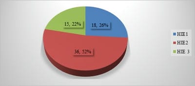
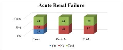
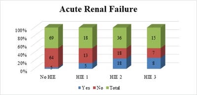
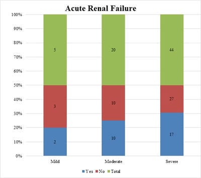
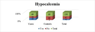
Background: Birth asphyxia is a common neonatal problem and contributes significantly to neonatal mortality and long term morbidity. Any organ can be affected but the brain, heart, kidneys are more sensitive to hypoxic injury. Aim and Objective: To study renal parameters and serum calcium levels in birth asphyxia, to determine the incidence of renal failure in asphyxiated newborns, and to correlate the renal parameters with different severity of birth asphyxia and with different stages of hypoxic-ischemic encephalopathy. Method and material: A comparative study of 69 term newborns, appropriate for gestational age born/admitted to tertiary care Centre RDGMC Ujjain with birth asphyxia and 69 normal newborns without birth asphyxia was done. Perinatal history, physical examinations, New Ballard score, Apgar score was done. Data collected and entered in the master chart for results and analysis. Result: The study included 69 cases and 69 controls with the majority of boys. Mild, moderate, and severe birth asphyxia was7%, 29%, and 64% of all cases respectively. The incidence of acute renal failure was 57% among cases of birth asphyxia. The incidence of acute renal failure was 53% and 50% in hypoxic-ischemic encephalopathy stages III and II respectively. Incidence of renal failure with mild, moderate, and severe asphyxia was 40%, 50%, and 39% respectively Conclusion: Perinatal asphyxia is an important cause of neonatal renal failure. Monitoring of urea, creatinine and urine output helps in the early diagnosis and management of acute renal failure

Keywords: Acute Renal failure, Birth asphyxia, Hypoxic Ischemic Encephalopathy (HIE), Sarnat and Sarnat Staging

Corresponding Author How to Cite this Article To Browse
Jagdish Chandra Mandliya, Professor, Department of Pediatrics, R.D. Gardi Medical College, Ujjain, Madhya Pradesh, India.
Email:
Patidar A, Mandliya JC, Sonker P, Dhaneria M. To study renal parameters and serum calcium levels in birth asphyxia. Pediatric Rev Int J Pediatr Res. 2020;7(6):255-261.
Available From
https://pediatrics.medresearch.in/index.php/ijpr/article/view/611

Manuscript Received Review Round 1 Review Round 2 Review Round 3 Accepted
2020-07-18 2020-07-31 2020-08-10 2020-08-25
Conflict of Interest Funding Ethical Approval Plagiarism X-checker Note
No Nil Yes 9%

© 2020 by Amit Patidar, Jagdish Chandra Mandliya, Pavan Sonker, Mamta Dhaneria and Published by Siddharth Health Research and Social Welfare Society. This is an Open Access article licensed under a Creative Commons Attribution 4.0 International License https://creativecommons.org/licenses/by/4.0/ unported [CC BY 4.0].

Introduction

The incidence of perinatal asphyxia is 1–1.5 % in most developed countries and is inversely related to gestational age and birth weight [1]. In India the incidence of perinatal hypoxia is as high as 8 -9% and it accounts for 28.8% of neonatal deaths and 45.1% of fresh still births [2].

According to the latest estimates by World Health Organization (WHO), approximately 4 million babies die each year before they reach the age of one month, ninety eight percent of these neonatal deaths take place in the developing countries.

WHO has defined perinatal asphyxia as a failure to initiate and sustain breathing at birth [3]. The National Neonatal Perinatal Database (NNPD) 2000 used a similar definition for perinatal asphyxia. It defined moderate asphyxia as slow gasping breathing or an Apgar score of 4-6 at 1 minute of age and severe asphyxia was defined as no breathing or an Apgar score of 0-3 at 1 minute of age [4]. As per the AAP (American Academy of Pediatrics) and ACOG (American College of Obstetrics and Gynecology), all the following must be present for designation of asphyxia

(a) Profound metabolic or mixed acidemia (pH< 7.00) in cord blood

(b) Persistence of Apgar score 0-3 for longer than 5minutes

(c) Neonatal neurologic sequelae (eg, seizures, coma, Hypotonia)

(d) Multiple organ involvement (kidney, lungs, liver, heart, intestine)

According to National Neonatal Perinatal Database (NNPD) 2003 data collected from 17 tertiary neonatal intensive care units in India, Apgar scores < 7 at 1 minute documented in 9% of all intramural delivery [5]. 2.5% of babies continued to have Apgar scores <7 at 5 minutes of age. Bag and mask ventilation was used in 4.5% newborn and less than 1% newborn needed cardiac compressions and/or medications for resuscitation at birth.

Perinatal asphyxia was responsible for 20% of all neonatal deaths. Manifestations of hypoxic-ischemic encephalopathy were seen in approximately 1.5% of all babies. Perinatal asphyxia was the commonest cause of still-birth accounting for one-third of all such cases [6].

Material and Method

Setting: NICU of tertiary care Centre R.D. Gardi Medical College Ujjain (M.P.)

Type and duration of study: Case-Control Study conducted from November 2017 to June 2019

Sampling Methods: Newborn with and without birth asphyxia were included in the study. At the time of enrollment, and informed written consent was obtained from the parents. Detailed perinatal history was obtained from hospital records. A detailed clinical examination was done. Newborn identified as appropriate for gestational age by applying the New Ballard score and by percentile chart. Diagnosis of birth asphyxia was made by Apgar score, Gestational age, birth weight, relevant perinatal history, findings on physical examination, and systemic examination.

Renal function parameters: Urine output, serum sodium, potassium, urea, creatinine, and calcium were sent to the biochemistry lab by taking peripheral venous blood samples, initially within 24hrs of birth. At 48hrs and 72hrs in addition, blood urea and creatinine were measured. Peripheral vein samples from all the neonates were collected in sterilized tubes and sent immediately to the Biochemistry lab for testing by – Dry chemistry/slide Method by Vitro’s -250 fully automatic analyzer.

Inclusion Criteria

For cases:

1. Term newborn with history of birth asphyxia

2. Evidence of neurological abnormalities suggestive of Hypoxic Ischemic Encephalopathy

3. The need for immediate neonatal resuscitation including bag and mask ventilation

4. Presence of immediate neonatal distress as evidenced by low Apgar score <7

For Control

1.The term, newborn who is appropriate for gestational age and normal birth weight (>2.5kg)

Exclusion Criteria

  1. Congenital abnormalities of kidneys and urinary tract.
  2. Babies with Septicemia, respiratory distress syndrome.

Procedures and plan: Renal parameters namely serum Urea, Serum Creatinine, Serum Sodium, serum Potassium, Calcium, Urine output measured within 24hrs of birth, 48hrs, and 72 hours of birth. Acute renal failure was defined as serum creatinine of >1.0mg/dl on day 3 of life or urine output <1.0ml/kg/hr. Hypocalcemia in the present study was defined as serum calcium level<8.0mg/dl

On the basis of Apgar score at 1minute asphyxiated babies grouped into mild (6) moderate (4-5) and Severe asphyxia ≤3 Apgar Score [7].

Neurological examination in the newborn at the time of admission and discharge was done by the following category:

  1. Level of alertness
  2. Cranial nerves
  3. Motor function
  4. Developmental reflexes
  5. Sensory response Gestational age assessment was done by using the New Ballard Score.

Collected data analyzed by unpaired and paired t-test, Karl Pearson’s Co-relation Co-efficient, and Chi-Square Test.

Peripheral venous samples from all the neonates were collected in sterilized tubes at the time of admission and sent immediately for analysis by the standard biochemical method.

Data Collection and Methods: Date observed and collected on individual cases entered in prerecorded proforma that entered in the master chart of observation for calculations, results, and analysis.

Data management and statistical analysis: Data was entered in Epidata entry (version3.1 Epidata Software Association, Odense, Denmark) and statistical analysis was performed using Stata (version 13.0, StataCorp, Texas, USA).

For continuous variables range, mean and standard deviation (SD) will be presented. Categorical independent variables are investigated using Pearson chi-square.

Logistic regression was employed to further investigate each independent variable with the dependent variable and odd’s ratios were derived. A p-value of <0.05 was regarded as significant

Ethical approval: The study was approved by the institutional ethical review board (IEC ref. no-172)

Results

Table 1: Sex distribution among cases and controls.

  Cases Control
Boys 48(70%) 44(64%)
Girls 21(30%) 25(36%)

The study included 69 cases and 69 controls with the majority of boys in both groups. There were 48boys among cases and 44 boys among controls

Table 2: Distribution of cases according to the severity of Asphyxia.

  Mild Moderate Severe
n (%) 5 (7%) 20 (29%) 44 (64%)

the study population includes the majority of neonates with moderate and severe birth asphyxia accounting for 29% and64% of all cases respectively. Mild asphyxia accounting for 7%

Table 3: Distribution of cases according to HIE staging.

HIE Stage n (%)
I 18 (26%)
II 36 (52%)
III 15 (22%)
Total 69 (100%)

pedia_611_01.jpg

Fig-1: Distribution of cases according to HIE staging.

Majority cases in HIE stage II were 52% and the remaining cases HIE stage III was 22% and HIE stage I was 26%

Table 4: Incidence of acute renal failure among cases and control.

  Cases Controls Total
Yes 39(57%) 0 (0%) 39
No 30(43%) 69 (100%) 99
Total 69 69 138

Chi2 = 38.5706 P = 0.000


pedia_611_02.jpg

Fig-2: Incidence of acute renal failure among cases and control.

Incidence of acute renal failure was 57% among cases which were statistically significant (P=0.000)

Table 5: Incidence of Acute Renal Failure in different stages of HIE.

  YES NO TOTAL Odds Ratio 95% CI P-value
No HIE 5 (7%) 64(93%) 69 R R R
HIE 1 5 (28%) 13 (72%) 18 32.28 8.41 0.00
HIE 2 18 (50%) 18 (50%) 36 12.8 4.17 0.00
HIE 3 8 (53%) 7 (47%) 15 14.62 3.74 0.00

Chi2 = 41.38 P = 0.000

pedia_611_03.jpg

Fig-3: Incidence of Acute Renal Failure in different stages of HIE.

The incidence of acute renal failure was 53% and 50% in HIE stages III and II respectively, and 28% of the neonates in stage I and with7% of No HIE group had renal failure.

The incidence of renal failure was highest in HIE stage III among cases with birth asphyxia. Incidence of acute renal failure in HIE stage I(P=0.000), stage II (P=0.000), stage III (P=0.000) was statistically significant

Table-6: Incidence of Renal Failure with the severity of asphyxia.

  Yes No Total Odds Ratio 95% CI P-Value
Mild 2 (40%) 3(60%) 5(7%) R R R
Moderate 10(50%) 10(50%) 20(29%) 1.5 0.204 -10.99 0.04
Severe 17(39%) 27(61%) 44(64%) 2.38 0.360 -15.75 0.036

Chi2 = 1.3213 P = 0.048

pedia_611_04.jpg

Fig-4: Incidence of Renal Failure with the severity of asphyxia.

The incidence of acute renal failure in mild, moderate, and severe asphyxia was 40%, 50%, and 39% respectively.

Incidence of renal failure in moderate (P=0.04) and severe asphyxia (P=0.036) was statistically significant

Table-7: Serum calcium levels among cases and controls.

S. Calcium Group Number Mean(mg/dl) Standard Deviation Z
< 24 Hours Cases 69 9.03 1.03 1.397
Controls 69 9.13 0.11 p = 0.163
48 Hours Cases 69 8.30 0.94 3.67
Controls 69 9.21 0.14 p < 0.001
72 Hours Cases 69 8.32 0.92 4.073
Controls 69 9.24 0.13 p < 0.001

In this study, the mean serum calcium levels in cases within 24 hours of life were 9.03 ± 1.03, at 48 hours of life, the mean value of serum calcium was 8.3 ± 0.94 with statistical significance and at 72 hours is 8.32 ± 0.92 also with statistically significant.

The study showed serum calcium levels were low among cases.

Table-8: Incidence of hypocalcemia among cases and controls.

Hypocalcemia Case Control Total
YES 40 (58%) 1 (2%) 41
NO 29 (42%) 68 (98%) 97
TOTAL 69 69 138

pedia_611_05.jpg

Fig-5: Incidence of hypocalcemia among cases and control.

Incidence of hypocalcemia was 58% in cases with birth asphyxia and 2% in control

Table-9: Incidence of hypocalcemia with the severity of asphyxia.

  Yes No Total Odds Ratio 95% CI P-value
Mild 1(20%) 4(80%) 5 R R R
Moderate 9(45%) 11(55%) 20 3.2727 0.3084 0.325
Severe 24(55%) 20(45%) 44 4.8 0.4957 0.04
Total 34 35 69      

Chi2 = 2.3496 P = 0.03

Incidence of hypocalcemia with mild, moderate, and severe asphyxia was 20%,45%, and 55% respectively. Incidence of hypocalcemia with moderate (P= 0.325) was statistically not significant and severe asphyxia (P=0.04) was statistically significant

Discussion

The incidence of acute renal failure in asphyxiated neonates with different stages of hypoxic-ischemic encephalopathy was determined, along with the incidence of acute renal failure with the severity of asphyxia.

Acute renal failure in the present study was defined as serum creatinine >1.0mg/dl on day3 of life [8].

The study done by Nilesh V. Ahire et al and colleague 2017 had defined acute renal failure as serum creatinine of level >1mg/dl on day 3 of life and/or urine output of <1ml/kg/hour [8]

Serum calcium levels in cases of birth asphyxia and control were calculated along with the incidence of hypocalcemia (S. Ca2+<8mg/dl) in cases of birth asphyxia [9].

The present study noted that boys were more common in cases and control both. It includes 70% boys among cases and 64% of boys among the control group out of 69 in each case and controls separately. In the study done by Mac Donald [10] and others, the incidence of asphyxia was 54% in

male babies and 46% in female babies which correlated well with the present study.

Ganavi et al and colleague (2016) concluded that the incidence of acute renal failure was significantly more in cases (75% vs 4%) and 18.4 times more likely when compared to controls.

In the present study, the incidence of acute renal failure was 57% among cases. The incidence of acute renal failure among cases (P=0.000) is statistically significant [9,11].

In the present study, the incidence of renal failure was comparable with the studies done by Gupta et al [12], Zulfikar Ali Mangi et al [13], Aggarwal et al [14] and Jayashree et al [15].

In the current study serum creatinine of more than 1 mg/dl after 72 hours was considered as renal failure. All studies showed that non-oliguric renal failure was most common in asphyxiated neonates.

The mean serum creatinine levels among the cases in the present study were 1.44+0.54 and 1.19+0.38 as compared to controls had 0.8+0.15 and 0.6+0.06 at 48 and 72 hours of life.

Table-10: Comparative studies showing mean serum creatinine levels among cases and controls.

Studies Year Cases ( mg/dL ) Controls
Gupta et al [12] 2005 1.08±0.49 0.88±0.26
Zulfikar ali mangi et al [13] 2009 1.95±0.45 0.83±0.17
Aggarwal et al [14] 2005 1.0 ±0.5 0.7±0.2

In the present study, it was found that serum creatinine levels were higher in cases as compared to controls and the difference between the groups was statistically significant both at 48 and 72 hrs.

But however, the serum creatinine value was higher in cases with severe asphyxia or with stage III HIE, the difference among the groups was not statistically significant.

In the present study the mean serum calcium value among the cases was 9.03±1.03 mg/dl (p=0.163), 8.30±0.94 (p<0.001) mg/dl, 8.32±0.92 mg/dl (p<0.001) at 24 hours, 48 hours and 72 hours respectively [9,16].

Serum calcium levels among Controls were 9.13±0.11mg/dl, 9.21±0.14mg/dl, 9.24±0.13mg/dl, at 24 hours, 48 hour and 72 hours [9,16].


Table-11: Comparative studies showing mean serum calcium levels among cases and controls.

Studies Year Cases (mg/dL) Controls (mg/dL)
Basu P et al [16] 2010 6.85 ± 0.95 9.50 + 0.51
Amrita et al [9] 2015 7.00 ± 0.691 8.788±3.91

Conclusion

The following conclusion was made in the present study:

  1. Male neonates had a higher incidence of birth asphyxia
  2. The incidence of renal failure had a linear correlation with the severity of asphyxia and with the HIE stage and incidence of intrinsic renal failure increased as the severity of asphyxia and HIE staging progressed. The creatinine was elevated among cases and had a strong correlation with severity of asphyxia and HIE staging
  3. The serum calcium was lower among cases and a significant difference was noted at 48 and 72 hours of life, the incidence of hypocalcemia was 58% among cases.
  4. Incidence of hypocalcemia was more among the case of birth asphyxia as compared to normal healthy newborn and there was a decrease in serum calcium levels with severity of asphyxia
  5. Perinatal asphyxia is an important cause of neonatal renal failure
  6. Monitoring of blood levels of urea, creatinine and urine output helps in the early diagnosis and management of renal failure
  7. In birth asphyxia, since non-oliguric renal failure is common monitoring only urine output does not help in the diagnosis of ARF, the biochemical parameters in both blood and urine should be monitored

What does the study add to the existing knowledge?

The renal indices should be calculated, as fractional excretion of sodium is preferred to classify the renal failure into pre-renal or intrinsic renal failure as management differs for both entities. Acute renal failure in birth asphyxia shows a strong positive correlation with HIE. In Hypoxic Ischemic Encephalopathy prevention intrinsic renal failure is

better than managing as it is associated with multi-organ dysfunction syndrome and has increased mortality.

Author’s contribution

Dr. Amit Patidar: Concept, study design

Dr. Jagdish Chandra Mandliya: Manuscript preparation

Dr. Pavan Sonker: Data analysis

Dr. Mamta Dhaneria: Manuscript preparation and data analysis

Reference

  1. Hansen AR, Soul JS. Perinatal asphyxia and Hypoxic ischemic encephalopathy in Manual of neonatal care. 7th ed India- Wolters Kluwer. 2011; 711-728;350-376.
  2. Ilah BG, Aminu MS, Musa A, Adelakun MB, Adeniji AO, Kolawole T. Prevalence and risk factors for perinatal asphyxia as seen at a specialist hospital in Gusau, Nigeria. Sub-Saharan Af J Med. 2015;2(2)64.
  3. World Health Organization. Perinatal mortality- a listing of available information. World Health Organization. 1996.
  4. Network NN. National Neonatal Perinatal Database–report for the year 2002–2003. NNF NNPD network; New Delhi. 2005.
  5. Agarwal R, Jain A, Deorari AK, Paul VK. Post-resuscitation management of asphyxiated neonates. Indian J Pediatr. 2008;75(2)175-180.
  6. Lawn J, Shibuya K, Stein C. No cry at birth- global estimates of intrapartum stillbirths and intrapartum-related neonatal deaths. Bulletin of the World Health Organization. 2005;83;409-417.
  7. Ahire NV, Patil SV, Patel ND, Sonawane R, Joshi D. A Comparative Study of Renal Parameters and Serum Calcium Levels in Birth Asphyxiated Neonates and Normal Neonates. MVP J Med Sci. 2017;4(2)97-101.

  1. Lawn JE, Cousens S, Bhutta ZA, Darmstadt GL, Martines J, Paul V, et al. Why are 4 million newborn babies dying each year?. The Lancet. 2004;364(9432)399-401.
  2. Vamne A, Thanna RC, Pathak S, Chavan N. A Study on Serum Calcium Level in Birth Asphyxia. Int J Health Sci Res. 2015;5(4)147-151.
  3. Mac Donald HM, Mulligan JC, Allen AC, Taylor PM. Neonatal asphyxia, Relationship of obstetric and neonatal complication to neonatal mortality in 38,405 consecutive deliveries. J Pediatr. 1980;96(5)898-902.
  4. Chetty S, Kajjam SK, Patel A. Incidence of acute renal failure in birth asphyxia and its correlation with hypoxic-ischemic encephalopathy staging. Indian J Child Health. 2017;387-389.
  5. Sarnat HB, Sarnat MS. Neonatal encephalopathy following fetal distress- A clinical and electroencephalographic study. Arch Neurol. 1976;33(10)695-706.
  1. Gupta BD, Sharma P, Bagla J, Parakh M, Soni JP. Renal failure in asphyxiated neonates. Indian Pediatr. 2005;42(9)928-934.
  2. Mangi ZA, Bouk GR, Junejo B, Junejo A, Shaikh SH, Jamro S. Birth asphyxia relationship between hypoxic ischemic encephalopathy grading and development of acute renal failure in indoor term neonates at chandka medical college children hospital Larkana. Pediatr. 2009;15;148-152.
  3. Aggarwal A, Kumar P, Chowdhay G, Mazumdar S, Narang A. Evaluation of renal functions in asphyxiated newborns. J Trop Pediatrics. 2005;51(5)295-299.
  4. Jayashree G, Dutta AK, Sarna MS, Saili A. Acute renal failure in asphyxiated Newborns. Indian Pediatr. 1991;28(1)19-23.
  5. Basu P, Som S, Das H, Chaudhuri N. Electrolyte status in birth asphyxia. Indian J Pediatr. 2010;77(3)259-262.