E-ISSN:2349-3267
P-ISSN:2349-5499

Research Article

Protein

Pediatric Review - International Journal of Pediatric Research

2021 Volume 8 Number 4 July-August
Publisherwww.medresearch.in

“Study of Estimation of Cerebrospinal Fluid C-Reactive Protein in Diagnosis of Acute Meningitis.”

Bansal K.1, Bhatt D.2*, Kumar Dadhich S.3, Kariya D.4
DOI: https://doi.org/10.17511/ijpr.2021.i04.02

1 Keshav Bansal, Senior Resident, Department of Pediatrics, Pacific Institute of Medical Science, Udaipur, Rajasthan, India.

2* Dhaval Bhatt, Assistant Professor, Department of Pediatrics, Government Medical College, Bhavnagar, Gujarat, India.

3 Sunil Kumar Dadhich, Assistant Professor, Department of Pediatrics, Government Medical College, Kota, Rajasthan, India.

4 Deep Kariya, PG Resident, Department of Pediatrics, Government Medical College, Bhavnagar, Gujarat, India.

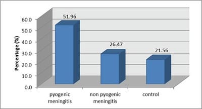
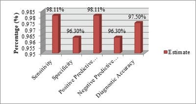
Objectives:To assess the diagnostic role of CSF C-reactive protein quantitatively in acute meningitis and to evaluate the efficacy of CSF C-reactive protein in differentiating pyogenic meningitis from non-pyogenic meningitis.Material and Methods: It is a prospective observational study of total 102 children with suspected meningitis allocated into three groups based on initial investigations; group-I Pyogenic meningitis, group-II Non-Pyogenic meningitis and group-III No meningitis (Control group). Quantitative CSF C-reactive protein was detected by the latex agglutination method. Data were analyzed to establish the diagnostic role of CSF-CRP and to evaluate the efficacy of CSF-CRP in differentiating pyogenic meningitis from non-pyogenic meningitis.Results: A total of 102 clinically suspected meningitis patients were studied. Based on CSF findings, the study population (102 cases) was categorized into 3 groups. Group I was pyogenic meningitis consist 53 cases (51.96%). Group II was Non-Pyogenic meningitis consists 27 cases (26.47%). Group III was normal CSF findings consist 22 (21.56%). 98.1% cases of pyogenic meningitis had elevated CSF-CRP level >1.1 µg/ml of CSF. In the case of Non-Pyogenic meningitis, 96.2% were found to have CSF- CRP in the range of 0.05-0.10 µg/ml. The mean value of CSF-CRP in groups I,II and III were 5.57±1.48, 0.09±0.042 and 0.01±0.010 respectively. Conclusion: Detection of CSF-CRP provides a new dimension to establish the diagnosis of pyogenic meningitis. It is a rapid, reliable and sensitive diagnostic test. From this study it is concluded that CSF-CRP can be used to differentiate pyogenic from non-pyogenic meningitis. Early, accurate and appropriate therapy can ameliorate the morbidity and mortality rates in such cases.

Keywords: CSF-CRP, Meningitis, Brain Infections

Corresponding Author How to Cite this Article To Browse
Dhaval Bhatt, Assistant Professor, Department of Pediatrics, Government Medical College, Bhavnagar, Gujarat, India.
Email:
Bansal K, Bhatt D, Dadhich SK, Kariya D. “Study of Estimation of Cerebrospinal Fluid C-Reactive Protein in Diagnosis of Acute Meningitis.”. Pediatric Rev Int J Pediatr Res. 2021;8(4):175-181.
Available From
https://pediatrics.medresearch.in/index.php/ijpr/article/view/660

Manuscript Received Review Round 1 Review Round 2 Review Round 3 Accepted
2021-03-03 2021-04-04 2021-05-26 2021-06-10 2021-07-05
Conflict of Interest Funding Ethical Approval Plagiarism X-checker Note
No Nil Yes 8%

© 2021 by Keshav Bansal, Dhaval Bhatt, Sunil Kumar Dadhich, Deep Kariya and Published by Siddharth Health Research and Social Welfare Society. This is an Open Access article licensed under a Creative Commons Attribution 4.0 International License https://creativecommons.org/licenses/by/4.0/ unported [CC BY 4.0].

Introduction

Acute infections of the nervous system are among the most important and common conditions in pediatrics. These distinct clinical syndromes include acute pyogenic meningitis, viral meningitis-encephalitis, focal infections such as brain abscess and subdural empyema, and infectious thrombophlebitis. Early recognition & diagnosis with the rapid institution of therapy can be life-saving[1].Meningitis is a significant cause of morbidity and mortality in children worldwide. Without treatment, the case-fatality rate can be as high as 70 percent, and one in five survivors of bacterial meningitis may be left with permanent sequelae including hearing loss, neurologic disability [2].

Case fatality rates for bacterial meningitis range from 4.5% in developed countries to 15–50% in developing countries [3,4]. Bacterial meningitis, still one of the most life-threatening problems worldwide, is more prevalent in children and its timely and early differentiation from non-bacterial meningitis has a huge impact on the treatment of affected patients. Quick diagnosis and effective management is key to success [5].

The evolution of clinical signs and symptoms caused by meningitis or encephalitis varies greatly. Thus, the clinician must sort out the form of clinical presentation, assess the rapidity of its evolution, and make a specific etiological diagnosis. The examination of cerebrospinal fluid is an essential and often critical tool in the evaluation and management of patients with meningitis. Although CSF examination is often useful for definitive diagnosis. But this is not always the case scenario. Cultures have the drawback of the time required, 24 to 48 hours or more to become positive, an unacceptable delay in initiating the treatment [6].Culture and sensitivity, Gram stain, cytology and biochemistry of cerebrospinal fluid (CSF) samples are traditionally being done to diagnose and to differentiate pyogenic from aseptic meningitis. CSF cytology and culture sensitivity reports may be affected by prior antibiotic therapy. Gram’s stain lacks specificity and has interpretative errors. Possible causes of the false-positive result of Gram’s stain include contamination of tubes from lumber puncture trays, glass slides and or Gram’s reagents. The overall sensitivity of Gram’s stain to detect bacterial meningitis was 67% with a positive predictive value of 60%.

Most patients without bacterial meningitis have a negative Gram’s stain (specificity 99.9%) with a negative predictive value of 99.9% [7].

Detection of nuclear polymorph leukocytes in the CSF is a fairly reliable indicator of pyogenic meningitis. But CSF leukocyte count < 250/mm3 may be present in as many as 20% of patients with bacterial meningitis. Pleocytosis may be absent in patients with severe overwhelming sepsis. Pleocytosis with a lymphocytic predominance may be present during the early stage of acute bacterial meningitis; conversely, neutrophilic pleocytosis may be present in patients during the early stages of acute viral meningitis Use of antibiotics makes the gram’s stain and culture-negative and may alter the CSF cytology from neutrophilic to lymphocytic predominance [8].

Because of these limitations, several rapid diagnostic tests have been developed to aid in the diagnosis& to discriminate rapidly between viral meningitis and bacterial meningitis [9]. These techniques include Counter Immuno Electrophoresis of the CSF for the immunoglobulins, lactic acid, creatine phosphokinase and C-reactive protein [10].As CRP is the fastest reacting and most sensitive indicator of an acute inflammatory reaction, it is a useful aid in preliminary differentiation between acute bacterial and non-bacterial meningitis. Detection of CSF-CRP appears to provide a new dimension to the diagnosis of meningitis [11].

Material and Methods

Place of Study: Department of Pediatrics at Government Medical College, Kota.

Type of study: Cross-Sectional, Descriptive, Observational Study.

Sampling Method: Convenient sampling

Sample Collection: Patients with suspected cases of meningitis with clinical signs and symptoms of acute meningitis, aged 1 month to 18 years, admitted to Department of Pediatrics, Government Medical College and Associated Group ofHospitals, Kota.

Inclusion criteria

  • Age 1 month to18 years.
  • Clinical features are suggestive of meningitis.
  • Patients with high body temperature.

  • Feeding problems.
  • Vomiting.
  • Irritability
  • Seizures or sluggishness.
  • High pitched crying

Exclusion criteria

  • Patients in whom lumbar puncture is contraindicated i.e.
  • Sepsis at the local site
  • Papilloedema or other signs of raised intracranial pressure
  • Marked spinal deformity
  • Bleeding diathesis or on anticoagulant therapy
  • Patients on steroid
  • Traumatic lumbar puncture
  • Refusal to consent
  • Patients having congenital CNS abnormality and who is known case of neurodegenerative disorder of the brain.

Statistical Method: ANOVA, unpaired t-test.

Sample Size: 102 patients were included after the protocol was approved by an ethical review committee of Government Medical College, Kota. Informed written consent was taken.A detailed history has been taken. A general physical and systemic examination was done. Investigations including Complete Blood Count with CSF analysis (appearance, cell count & differential, sugar, protein, gram’s stain, culture), quantitative CSF-CRP, blood sugar, Mantoux Test (MT) in tubercular suspected, cranial CT scan and MRI brain if indicated was done.

Patients investigated as above were divided into three groups as mentioned below:

Group-I: Pyogenic meningitis

Group-II: Non-Pyogenic meningitis (Tubercular meningitis, Aseptic meningitis, etc.)

Group-III:- No Meningitis (Normal CSF: cerebral malaria, febrile convulsion, dyselectrolytemia etc.)

We assessed the role of CSF C-reactive protein quantitatively in acute meningitis by using compute based program statistical package for social science (SPSS) version 20.0 programs.

Results

Total 102 clinically suspected meningitis children were enrolled for this cross-sectional observational study.

Among 102 cases, 53 (51.96%) were diagnosed as Pyogenic meningitis (Group I), 27 (26.47%) as Non-Pyogenic meningitis (Group II) and 22 (21.56%) as No meningitis (Group III, normal CSF).[Figure 1]

Figure 1: Categories of the study population based on CSF findings

pedia_660_01.jpg

Figure 2: CSF-CRP Test Estimation

pedia_660_02.jpg

In most of the cases 65 (63.6%) of the study population were in the age group of 1 month to 5 years. The age distribution among Pyogenic meningitis (53) shows the maximum of 17 cases (32.0%) in the age group 1 month to 1 year followed by 15 (28.3%) in the age of >1 year to 5 years [Table 1]. Sex distribution of study population where male patients were 58 (56.8%) and female 44 (43.1%).

However, the difference between males and females was not statistically significant [Table2]. Clinical findings of acute meningitis in the study population where all of the study population (102) were suffering from fever (100%), Headache were present in Pyogenic meningitis 16 (30.1%) and Non-Pyogenicmeningitis 7 (25.9%) respectively. Other clinical findings of enrolled children were described in Table 3.


Table- 1: Age distribution of the study population (N=102)

Age group Pyogenic meningitis Non-Pyogenic meningitis Normal CSF (Controls) Total
1 month to 1 year 17 (32.0) 5 (18.5) 4 (18.1) 26 (25.4%)
>1year to 5 years 15 (28.3) 12 (44.4) 12 (54.5) 39 (38.2%)
>5 years to 10 year 14 (26.4) 4 (14.8) 5 (22.7) 23 (22.5%)
>10 years to 18 years 7 (13.2) 6 (22.2) 1 (4.5) 14 (13.7%)
Total 53 (100%) 27 (100%) 22 (100%) 102 (100%)

Table-2: Sex distribution of the study population (N=102)

Sex Pyogenic meningitis Non-Pyogenic meningitis Normal CSF (Controls) Total
Male 31 (58.4) 15 (55.5) 12 (54.5) 58 (56.8)
Female 22 (41.5) 12 (44.4) 10 (45.4) 44 (43.1)
Total 53 (100%) 27 (100%) 22 (100%) 102 (100%)

Table 3: Clinical Presentation

Clinical findings Pyogenic meningitis (Group I) Non-Pyogenic meningitis (Group III) Normal CSF (Controls) (Group III) Total (N=102)
Fever 53 (100%) 27 (100%) 22 (100%) 102 (100%)
Headache 16 (30.1%) 7 (25.9%) 2 (9%) 25 (24.5)
Nausea/ Vomiting 36 (67.9%) 18 (66.6%) 13 (59%) 67 (65.6%)
Altered Sensorium 46 (86.7%) 24 (88.8%) 13 (59%) 83 (81.3%)
Convulsion 29 (54.7%) 13 (48.1%) 22 (100%) 64 (62.7%)
Neck Rigidity 28 (52.8% ) 16 (59.2%) 0 44 (43.1%)
Kerning’ Sign 21 (39.6%) 8 (29.6%) 0 29 (28.4%)
Brudzinski’ Sign 17 (32%) 9 (33.3%) 0 26 (25.4%)

Cytological examination shows 41 (77.1%) cases of pyogenic meningitis had WBC count in the range of 100 to 2000 cell/mm3. In Non-Pyogenicmeningitis, where 26 (96.2%) were in the range of 6 to 500 cell/mm3. The mean value of the total count of WBC/mm3 in CSF, in pyogenic meningitis was 910.15+ 808.76, in Non-Pyogenic meningitis was 154.37+139.22 and in the case of normal CSF was 1.86+1.58.All 53 cases (100%) of pyogenic meningitis had more than 50%Polymorphs in CSF. In Non-Pyogenicmeningitis where 27 cases (100%) were cell type is Lymphocytes was > 50%. The mean value of Polymorphs in CSF, in pyogenic meningitis was 74.09+10.47, in Non-Pyogenic meningitis was 75.29+9.01 and in the case of normal CSF was 68.18+47.67.

All 53 (100.0%) cases of Pyogenic meningitis had low glucose level <90 mg/dl of CSF which is less than two-third of blood glucose level. In the case of Non-Pyogenic meningitis 9 (33.3%) were found to have in the range of 45 to 55 mg/dl. The mean value of Glucose level (mg/dl) in CSF, in pyogenic meningitis was 40.50±15.39, in Non-Pyogenic meningitis was 64±18.62 and in the case of normal CSF was 64.81±5.58. All 53 (100.0%) cases of Pyogenic meningitis had elevated protein levels>45 mg/dl of CSF. In the case of Non-Pyogenic meningitis 17 cases (62.9%) were found to have in the range of 46-400 mg/dl. The mean value of protein level (mg/dl) in CSF, in Pyogenic meningitis was 141.37±56.41, in Non-Pyogenic meningitis was 85.18±61.65 and in the case of normal CSF was 64.81± 5.58[Table 4].


Table- 4: Comparison of cytological and biochemical examination of CSF of the study population (N=102)

Parameters Pyogenic meningitis (n=53) Non-Pyogenic meningitis (n=27) Normal CSF (Controls) (n=22) P value*  
Cell Count (cells/cmm) 910.15+ 808.76 154.37+139.22 1.86+1.58 0.0001
Cell_type   74.09+10.47 (Polymorphs) 75.29+9.01 (Lymphocytes) 68.18+47.67 (Lymphocytes) 0.0001
CSF Sugar (mg/dl) 40.50+15.39 64+18.62 64.81+5.58 0.0001
CSF Protein (mg/dl) 141.37+56.41 85.18+61.65 64.81+5.58 0.0001
CSF CRP (µg/ml) 5.57+1.48 0.09+0.042 0.01+0.010 0.0001

In 52 cases (98.1%) of Pyogenic meningitis had elevated CSF-CRP level >1.1µg/ml and 1 case (1.8%) were in the range of 0.05-0.10 µg/ml. In the case of Non-Pyogenic meningitis 27 (96.2%) were found to have in the range of 0.05-0.10 µg/ml.

The mean value of CSF CRP level in Pyogenic meningitis was 5.57±1.48 µg/ml, in Non-Pyogenic meningitis was 0.09±0.042 and in case of normal CSF was 0.01±0.010 [Table 5, 6].


Table- 5: CSF-CRP level among the study population (N=102)

CSF CRP (µg/ml) Pyogenic meningitis (n=53) Mean ±SD Non-Pyogenic meningitis (n=27) Mean ±SD Normal CSF (Controls) (n=22) Mean ±SD
0.001-0.04 0 (0.0%) 5.57±1.48 0 (0.0%) 0.09±0.042 22 (100%) 0.01±0.010
0.05-0.10 1 (1.8%) 26 (96.2%) 0 (0.0%)
0.11-1 0 (0.0%) 1 (3.7%) 0 (0.0%)
1.1-15 52 (98.1%) 0 (0.0%) 0 (0.0%)

Table-6: CSF-CRP Test Evaluation

CSF CRP (µg/ml) Pyogenic meningitis (n=53) Non-Pyogenic meningitis (n=27) Normal CSF (Controls) (n=22) Total
more than 0.1ug/ml 52 1 0 53
less than 0.1ug/ml 1 26 22 49
Total 53 27 22 102

There was a highly significant difference in the total count of WBC, the level of Glucose and protein in CSF among Pyogenic meningitis, Non-Pyogenic meningitis and normal CSF (p-value < 0.05) [Table 7].

Table- 7: Multiple comparisons

Dependent Variable Mean Difference Std. Error P value
Cell Count(cells/cmm) Pyogenic meningitis Vs Non-Pyogenic meningitis 755.78 139.61156 0.0001
Controls 908.28 149.75370 0.0001
Non-Pyogenic meningitis Vs Pyogenic -755.78 139.61156 0.0001
Controls 152.50 169.59031 0.642
CSF Sugar (mg/dl) Pyogenic meningitis Vs Non-Pyogenic meningitis -23.49 3.52504 0.0001
Controls -24.30 3.78111 0.0001
Non-Pyogenic meningitis Vs Pyogenic 23.49 3.52504 0.0001
Controls -.818 4.28197 0.980
CSF Protein (mg/dl) Pyogenic meningitis Vs Non-Pyogenic meningitis 56.19 12.22859 0.0001
Controls 109.96 13.11694 0.0001
Non-Pyogenic meningitis Vs Pyogenic -56.19 12.22859 0.0001
Controls 53.77 14.85443 0.001
CSF CRP (µg/ml) Pyogenic meningitis Vs Non-Pyogenic meningitis 755.7 139.61 0.0001
Controls 908.28 149.75 0.0001
Non-Pyogenic meningitis Vs Pyogenic -755.78 139.61 0.0001
Controls 152.50 126.37 0.642

* The mean difference is significant at the 0.05 level.

Table 8 shows that in the pyogenic meningitis group, CSF culture was positive only in 15% of patients and common bacteria were grown in CSF culture were Streptococcus pneumoniae (7.5%), Staphylococcus aureus (3.7%) and H. influenza (1.8%) and E.Coli (1.8). Among the 4 cases of Streptococcus pneumoniae meningitis, 2 cases were below 12 months and 2 cases were more than 12 months (48 months& 6year).

Table 8: Bacteriology in Pyogenic meningitis.

Bacteriology No. of cases (n=53)
Streptococci 4 (7.5%)
H. influenza 1 (1.8%)
Staphylococcus aureus 2 (3.7%)
E. coli 1 (1.8%)
Culture positive 8 (15%)

Table 9 and Figure 2 Showing CSF CRP value of >0.1 μg/ml has a good sensitivity (98.11%), specificity (96.3%), good positive predictive value


(98.11%), and good negative predictive value (96.3%) and good Diagnostic Accuracy (97.5%) for Pyogenic meningitis.

Table 9: CSF-CRP Test Estimation

Parameter Estimate
Sensitivity 98.11%
Specificity 96.30%
Positive Predictive Value 98.11%
Negative Predictive Value 96.30%
Diagnostic Accuracy 97.50%
Positive Likelihood Ratio 26.49
Negative Likelihood Ratio 0.02

Discussion

C-reactive protein (CRP), an acute-phase inflammatory response reactantwas discovered in 1930 by Tillett et al [12].CRP is the acute phase protein that is synthesized by hepatocytes in response to infection or inflammation. In healthy the median concentration of serum C - reactive protein is 0.8mg/l. CRP concentrations in the CSF is much lower than those of serum. Meningeal irritation stimulates C-reactive protein production. Its production is controlled by Interleukin-6 and it binds to polysaccharides present in many bacteria, fungi and protozoal, parasites [13].Almost any inflammatory disease will cause detectable quantities of CRP to be present in serum or fluids closely associated with affected tissues [14,5,16].

Increased CRP production is an early and sensitive response to most forms of microbial infections and the value of its measurement in the diagnosis and management of various infective conditions has been established [17,18].In western countries, attention was recently drawn to the value of serum CRP measurement in differentiating bacterial and non-bacterial infections [19].However, routine diagnostic use of CSF- CRP in differentiating bacterial and non-bacterial meningitis has been evaluated by very few workers. Since meningitis is a potentially hazardous disease of childhood, diagnostic test which is readily available, easy to interpret and simple to perform is of paramount importance.

This report demonstrates the usefulness of a latex agglutination test for the detection of CSF-CRP as a rapid and simple, method in diagnosing and differentiating cases of bacterial and non-bacterial meningitis in children [20]. The concentration of CSF CRP is raised in patients with meningitis.

CSF-CRP has been reported to be one of the most reliable and early indices to differentiate bacterial from non-bacterial meningitis [21]. CRP estimation can help in diagnosing cases of acute bacterial meningitis more effectively than culture.It is also useful in monitoring the clinical course of meningitis [22]. This study aids to evaluate the diagnostic significance of CSF-CRP and as an indicator in the differentiation of bacterial from non-bacterial meningitis.

Conclusion

In conclusion we found that CSF-CRP level is significantly higher in pyogenic meningitis compared to non-pyogenic meningitis patients. It is a good indicator to diagnose pyogenic meningitis. It can be used to differentiate pyogenic from non-pyogenic meningitis also.  It is recommended to detect CSF-CRP level as a rapid diagnostic test to start appropriate antibiotic therapy, As Early, accurate and appropriate therapy can ameliorate the mortality and morbidity rates, the overall cost of the treatment and the duration of hospitalization.

Contributions details

Data collection was done by Dr. Keshav Bansal.

Analysis and manuscript preparation was done by Dr. Dhaval Bhatt and Dr. Deep Kariya.

All research works had been done under the guidance of Dr. Sunil Kumar Dadhich.

Reference

01. Karen L Roos, Longo, Fauci,Kasper et al. Pyogenic meningitis, In- Kenneth L Tyler, editor, Harrison’s principles of internal medicine. 18th ed, United States Of America- McGraw Hill. 2012;3410–20. [Crossref][PubMed][Google Scholar]

02. Rosenstein NE, Perkins BA, Stephens DS, Popovic T, Hughes JM. Meningococcal disease. N Engl J Med. 2001 May 3;344(18)1378-88. doi: 10.1056/NEJM200105033441807 [Crossref][PubMed][Google Scholar]

03. Baraff LJ, Lee SI, Schriger DL. Outcomes of bacterial meningitis in children- a meta-analysis. Pediatr Infect Dis J. 1993 May;12(5)389-94. doi: 10.1097/00006454-199305000-00008 [Crossref][PubMed][Google Scholar]


04. Salih MA, el Hag AI, Sid Ahmed H, Bushara M, Yasin I, Omer MI, et al. Endemic bacterial meningitis in Sudanese children- aetiology, clinical findings, treatment and short-term outcome. Ann Trop Pae diatr. 1990;10(2)203-10. doi: 10.1080/02724936. 1990.11747431[Crossref][PubMed][Google Scholar]

05. Salih MA, Khaleefa OH, Bushara M, Taha ZB, Musa ZA, Kamil I, et al. Long term sequelae of childhood acute bacterial meningitis in a developing country- A study from the Sudan. Scand J Infect Dis. 1991;23(2)175-82. doi: 10.3109/00365 549109023397 [Crossref][PubMed][Google Scholar]

06. Deivanayagam N, Ashok TP, Nedunchelian K, Ahamed SS, Mala N. Evaluation of CSF variables as a diagnostic test for bacterial meningitis. J Trop Pediatr. 1993 Oct;39(5)284-7. doi: 10.1093/tropej/39.5.284 [Crossref][PubMed][Google Scholar]

07. Neuman MI, Tolford S, Harper MB. Test characteristics and interpretation of cerebrospinal fluid gram stain in children. Pediatr Infect Dis J. 2008 Apr;27(4)309-13. doi: 10.1097/INF.b013e 31815f53ba [Crossref][PubMed][Google Scholar]

08. Whittle HC, Tugwell P, Egler LJ, Greenwood BM. Rapid bacteriological diagnosis of pyogenic meningitis by latex agglutination. Lancet. 1974 Sep 14;2(7881)619-21. doi: 10.1016/s0140-6736(74)91943-6 [Crossref][PubMed][Google Scholar]

09. Chowdhury MZU, Rahman KM, Miah RA, SattarH, Hussain T. Bacterial Meningitis in children. Bangladesh Medical Journal. 1991;3–7. [Crossref][PubMed][Google Scholar]

10. Brown RL, Zinner SH, Meglio FD, Garrity FL. Countercurrent immunoelectrophoresis in the diagnosis of viral infections of the central nervous system. J Infect Dis. 1978 Dec;138(6)911-5. doi: 10.1093/infdis/138.6.911 [Crossref][PubMed][Google Scholar]

11. Pemde HK, Harish K, Thawrani YP, Shrivastava S, Belapurkar KM. C-reactive protein in childhood meningitides. Indian J Pediatr. 1996 Jan-Feb;63(1)73-7. doi: 10.1007/BF02823870 [Crossref][PubMed][Google Scholar]

12. Tillett ws, francis t. Serological reactions in pneumonia with a non-protein somatic fraction of pneumococcus. J exp med. 1930 sep 30;52(4)561-71. Doi: 10.1084/jem.52.4.561 [Crossref][PubMed][Google Scholar]

13. Pepys MB, Hirschfield GM. C-reactive protein: a critical update. J Clin Invest. 2003 Jun;111(12):1805-12. doi: 10.1172/JCI18921 [Crossref][PubMed][Google Scholar]

14. Corrall CJ, Pepple JM, Moxon ER, Hughes WT. C-reactive protein in spinal fluid of children with meningitis. J Pediatr. 1981 Sep;99(3):365-9. doi: 10.1016/s0022-3476(81)80319-8 [Crossref][PubMed][Google Scholar]

15. Horsfall F L Jr, Tamm I. "Viral and Rickettsial Infections of Man". J B Lippincott Company, Philadelphia. 1965;pp-1167–8. [Crossref][PubMed][Google Scholar]

16. Pepys MB. C-reactive protein fifty years on. Lancet. 1981 Mar 21;1(8221):653-7. doi: 10.1016/s0140-6736(81)91565-8 [Crossref][PubMed][Google Scholar]

17. de Beer FC, Kirsten GF, Gie RP, Beyers N, Strachan AF. Value of C reactive protein measurement in tuberculous, bacterial, and viral meningitis. Arch Dis Child. 1984 Jul;59(7):653-6. doi: 10.1136/adc.59.7.653 [Crossref][PubMed][Google Scholar]

18. McCarthy PL, Frank AL, Ablow RC, Masters SJ, Dolan TF Jr. Value of the C-reactive protein test in the differentiation of bacterial and viral pneumonia. J Pediatr. 1978 Mar;92(3):454-6. doi: 10.1016/s0022-3476(78)80448-x [Crossref][PubMed][Google Scholar]

19. Peltola HO. C-reactive protein for rapid monitoring of infections of the central nervous system. Lancet. 1982 May 1;1(8279):980-2. doi: 10.1016/s0140-6736(82)91989-4 [Crossref][PubMed][Google Scholar]

20. Clarke D, Cost K. Use of serum C-reactive protein in differentiating septic from aseptic meningitis in children. J Pediatr. 1983 May;102(5):718-20. doi: 10.1016/s0022-3476(83)80242-x [Crossref][PubMed][Google Scholar]

21. Vaidya AK, Wagle NM, Merchant SM. Use of CSF C-reactive protein in differentiating bacterial and non-bacterial meningitis. J Postgrad Med. 1987 Apr;33(2):58-60. [Crossref][PubMed][Google Scholar]

22. Singh N, Arora S, Kahlon PS. Cerebrospinal fluid C-reactive protein in meningitis. Indian Pediatr. 1995 Jun;32(6):687-8. [Crossref][PubMed][Google Scholar]