E-ISSN:2349-3267
P-ISSN:2349-5499

Case Report

Disorder

Pediatric Review - International Journal of Pediatric Research

2021 Volume 8 Number 2 March-April
Publisherwww.medresearch.in

Tracheal agenesis: a rare congenital disorder

Gundapuneni Rao R.1, Susarla Venkata Rama B.2*, Gattu H.3, Paripati A.4
DOI: https://doi.org/10.17511/ijpr.2021.i02.01

1 Ravali Gundapuneni Rao, Fellow in Neonatology, Department of Neonatal Intensive care unit, Ankura hospital for women and children, Hyderabad, Telangana, India.

2* Balaji Susarla Venkata Rama, Consultant Neonatologist, Department of Neonatal Intensive care unit, Ankura hospital for women and children, Hyderabad, Telangana, India.

3 Harshitha Gattu, Consultant Neonatologist, Department of Neonatal Intensive care unit, Ankura hospital for women and children, Hyderabad, Telangana, India.

4 Alekya Paripati, Junior Medical Officer, Department of Neonatal Intensive care unit, Ankura hospital for women and children, Hyderabad, Telangana, India.

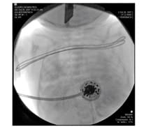
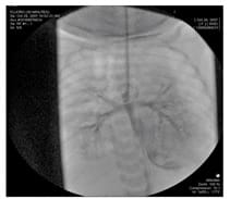
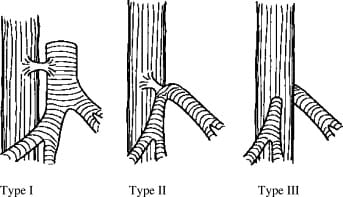
Tracheal agenesis is a severe congenital disorder with often an emergency presentation. There is a complete or partial absence of the trachea below the larynx, with the presence or absence of a tracheoesophageal fistula. It is a rare differential diagnosis of postnatal respiratory distress and the obstetrician or neonatologist will regularly be surprised by this malformation. The etiology of Tracheal atresia is unknown, therapeutic options are limited thus making this condition a usually fatal disorder. In most cases, congenital anomalies of the heart, digestive tract or GUT are present. The main signs are respiratory distress and cyanosis, inability to vocalize and impossible tracheal intubation. Isolated tracheal agenesis without any malformation is very rare. Here we report a case of a preterm neonate born at 33 weeks gestation with tracheal agenesis with no other associated malformation. In most cases, the seldom deformation, tracheal agenesis does not get recognized before the child is born.

Keywords: Tracheal agenesis, Congenital airway obstruction, Delivery room respiratory emergency

Corresponding Author How to Cite this Article To Browse
Balaji Susarla Venkata Rama, Consultant Neonatologist, Department of Neonatal Intensive care unit, Ankura hospital for women and children, Hyderabad, Telangana, India.
Email:
Rao RG, Rama BSV, Gattu H, Paripati A. Tracheal agenesis: a rare congenital disorder. Pediatric Rev Int J Pediatr Res. 2021;8(2):68-71.
Available From
https://pediatrics.medresearch.in/index.php/ijpr/article/view/663

Manuscript Received Review Round 1 Review Round 2 Review Round 3 Accepted
2021-03-06 2021-03-16 2021-03-24 2021-03-27
Conflict of Interest Funding Ethical Approval Plagiarism X-checker Note
No Nil Yes 5%

© 2021 by Ravali Gundapuneni Rao, Balaji Susarla Venkata Rama, Harshitha Gattu, Alekya Paripati and Published by Siddharth Health Research and Social Welfare Society. This is an Open Access article licensed under a Creative Commons Attribution 4.0 International License https://creativecommons.org/licenses/by/4.0/ unported [CC BY 4.0].

Introduction

Some congenital anomalies are not amenable to surgical treatment. Tracheal agenesis is one such congenital anomaly with limited therapeutic options. This lethal defect has an incidence of 2 per 1,00,000 live births [1]. It was first described by Payne in 1900 and later classified into 3 types by Floyd[2,3]. So far close to 200 cases have been reported in the literature with only a handful of cases reporting long-term survival [4]. It is frequently combined with other anomalies of the vertebrae, anal atresia, cardiovascular anomalies, tracheoesophageal atresia, renal/radial anomalies and limb defects(VACTERL)and tracheal agenesis, cardiac, renal and duodenal malformation (TACARD association) [5]. We report a case of antenatally unknown tracheal agenesis with no other major abnormalities and reviewed the literature.

Case report

A male infant was born to a 23-year-old primi mother by cesarean section. There was no history of consanguinity. Pregnancy was complicated by polyhydramnios but a routine antenatal ultrasound did not pick up any abnormality. The baby was born at 33 weeks of gestation because of the preterm onset of labor and fetal bradycardia. The infant was severely depressed at birth with a 1-min Apgar score of 2. There was no audible cry. The birth weight of the baby was 1750 grams. Positive pressure ventilation was started with a bag and mask, but there was no adequate chest rise. Direct laryngoscopy revealed a visible glottic plane with unaffected vocal cord mobility, but it was impossible to advance tracheal tubes of various sizes beyond the vocal cords.

Examination showed a hypoxic newborn with a PO2 of 57, with a blood pH of 6.9 and a PCO2 of 112. Mask ventilation was also difficult, but an O2 saturation of 87-90% was achieved following the administration of 75% FiO2. Chest X-ray was done which didn’t show any tracheal shadow- likely possibility of tracheal atresia. Esophageal intubation showed an improvement in saturations, suggesting communication between the respiratory and gastrointestinal tract.

An urgent referral was made to the otorhinolaryngologist. Once stabilized, the perinatal patient was immediately transferred to the operating room of a tertiary pediatric medical centre.

Repeat intubation using a 2.5 mm Endotracheal tube failed due to subglottic obstruction. Rigid bronchoscopy was performed and revealed a blind pouch below the vocal folds (Fig 1.)

pedia_663_01.jpg

Fig 1: The subglottic segment is markedly narrowed demonstrating absent cervical and thoracic trachea.

Intraoperative fluoroscopy of the trachea also confirmed the subglottic stenosis and absence of the lower cervical and thoracic trachea. The presence of a bronchoesophageal fistula was identified with both bronchi originating from a flattened carina. The bronchi and carina exhibited unusual horizontal orientation and the carina demonstrated a straight continuous roof with no tracheal remnants (Fig.2).

pedia_663_02.jpg

Fig 2: Note the horizontal orientation of the right and left bronchus and the carina.

Based on these findings, type II Tracheal atresia with bronchioesophageal fistula was diagnosed.


The patient died within 6 hours of birth. Parents were not willing for autopsy.

Discussion

Tracheal agenesis is a rare congenital defect that confronts Obstetricians, Neonatologists and Anesthesiologists with an emergency presentation. The diagnosis should be suspected in newborns with respiratory distress whose intubation is impossible. This condition has a male predominance and is often associated with prematurity and polyhydramnios [3,6]. The etiology remains unknown, although several developmental theories have been proposed. TA (Tracheal agenesis) is best explained by the recent theory that states that the lower respiratory tract develops as a respiratory diverticulum from the ventral aspect of the foregut which then elongates caudally to form the trachea. This is different from the earlier accepted theory that caudocranial fusion of the lateral ridges results in the division of the foregut into the ventral trachea and dorsal esophagus. The new hypothesis is based on an analysis of anomalous Tracheal oesophagal development in doxorubicin-exposed rat embryos. TA occurs with the development of a ventral rather than a dorsal pouch from the upper foregut, resulting in an atretic proximal trachea, while dorsal to that the foregut differentiates into the esophagus [7,8].

Tracheal agenesis is usually classified according to Floyd into three types (Fig. 1)3. In type I (10%) the trachea is absent, except for a short caudal part, and there is a normal carina. In type II (59 %) the complete trachea is lacking but both main bronchi join in the midline. In type III (31%) both the main bronchi run separately into the oesophagus; these connections are often referred to as broncho- oesophagal fistula [3]. Almost invariably the esophagus communicates with the trachea or bronchi; only in type II, a few exceptional cases without a fistula been described [2,9].

pedia_663_03.jpg

Fig 3. Classification of tracheal atresia by Floyd.

In our case, as demonstrated by rigid bronchoscopy and fluoroscopy, it was a case of tracheal atresia with both bronchi and carina having an unusual horizontal orientation with carina demonstrating a straight continuous roof with no obvious tracheal remnants.

The majority of TA (90%) is associated with multiple congenital anomalies involving the cardiovascular, gastrointestinal and genitourinary systems, making this condition usually uncorrectable and incompatible with life [10]. In our case, the baby did not have any obvious associated external congenital anomalies.

.The management of neonates with tracheal agenesis is very difficult. Any definitive therapy will have to be surgical. However, to date it has not been possible to do anything more than to provide a temporary airway. Different surgical reconstructive procedures have been developed, using either the oesophagus or synthetic material to create an upper airway. None of them has turned out to be effective concerning the long-term survival of the patients. Most of the patients died within a few hours to a few days [11,12]. In the future, generating a new tracheal tissue utilizing tissue engineering might be an approach to repair tracheal defects like tracheal atresia of tracheal agenesis. Animal experiments showed promising results in this direction [13].

Conclusion

Postnatal diagnosis of Tracheal agenesis is usually a respiratory emergency. The clinical features become manifest immediately after birth and are: (I) serious respiratory distress and cyanosis; (2) inability to vocalize despite obvious efforts; (3) on direct laryngoscopy the larynx often looks normal, but intubation fails due to an impassable resistance just beyond the vocal cords; (4) administration of oxygen with bag and masks under positive end-expiratory pressure can result in an improvement, but will often be associated with distension of the stomach.

Mask ventilation via the esophagus and the fistula may improve the condition of the neonate. Esophageal intubation and PEEP ventilation are approaches to secure a patent upper airway. Postnatal management of tracheal agenesis is a respiratory emergency and no obvious definitive therapy exists to date.


Antenatal diagnosis of the condition provides an early warning and greater preparedness to handle the situation.. However, our case was not antenatally diagnosed and the infant’s condition deteriorated rapidly despite possibly esophageal intubation with bag-mask ventilation.

This case report highlights that a potentially lethal anomaly like TA can exist in a presumably uncomplicated pregnancy. The presence of respiratory distress with an inaudible cry and inability to intubate should raise suspicion of this condition. Proper counseling of the parents is needed immediately after birth to prepare the parents for this potentially lethal condition.

Reference

  1. Manschot HJ, Anker JN, Tibboel D. Tracheal agenesis. Anaesthesia. 1994;49;788–790.
  2. Payne WA. Congenital absence of the trachea. Brooklyn Medical Journal. 1900;14;568.
  3. Floyd J, Campbell FJ, Dominy DE. Agenesis of the trachea. Am Rev Respir Dis. 1962;86;557–560.
  4. Mohammed H, West K, Bewick J, Wickstead M. Tracheal agenesis, a frightening scenario. J Laryngol Otol. 2016 Mar;130(3)314-7.
  5. Krause U, Rödel RM, Paul T. Isolated congenital tracheal stenosis in a preterm newborn. Eur J Pediatr. 2011;170(9)1217-1221.
  6. Hirakawa H, Ueno S, Yokoyama S et al. Tracheal agenesis- A Case Report. Tokai J Exp Clin Med. 2002;27;1-7.
  7. Merei JM, Hutson JM. Embryogenesis of tracheoesophageal anomalies- a review. Pediatr Surg Int. 2002;18;319–26.
  8. Strouse PJ, Newman B, Hernandez RJ, et al. CT of tracheal agenesis. Pediatr Radiol. 2006;36;920–6.
  1. Milles G, Dorsey DB. Intra-uterine respiration-like movements in relation to development of the fetal vascular system. American Journal of Pathology. 1950;26(41)1-25.
  2. Iszuari M, Mazita A, Tan GC, Hayati AR, Shareena I, Cheah FC. Tracheal agenesis- a rare cause of unsuccessful tracheal intubation during resuscitation. Med J Malaysia. 2010 Dec;65(4)317-8.
  3. Coleman AM, Merrow AC, Elluru RG, Polzin WJ, Lim FY. Tracheal agenesis with tracheoesophageal fistulae- fetal MRI diagnosis with confirmation by ultrasound during an ex utero intrapartum therapy (EXIT) delivery and post delivery MRI. Pediatr Radiol. 2013 Oct;43(10)1385-90.
  4. Kerschner J, Klotch DW. Tracheal agenesis- a case report and review of the literature. Otolaryngol Head Neck Surg. 1997 Jan;116(1)123-8.
  5. Veenendaal MB, Liem KD, Marres HA. Congenital absence of the trachea. Eur J Pediatr. 2000;159;8.