E-ISSN:2349-3267
P-ISSN:2349-5499

Case Report

Muscular

Pediatric Review - International Journal of Pediatric Research

2021 Volume 8 Number 3 May-June
Publisherwww.medresearch.in

Closure of a large upper muscular ventricular septal defect in a 2.3 kg baby using a duct occluder

Mohd Kamran M.1*, Abqari S.2, Haseen A.3, Yadav M.4
DOI: https://doi.org/10.17511/ijpr.2021.i03.07

1* Mirza Mohd Kamran, Assistant professor, Department of Pediatric Cardiology, PCE CS unit, JNMCH, AMU, Aligarh, Uttar Pradesh, India.

2 Shaad Abqari, Associate Professor, Department of Pediatrics, JNMCH, AMU, Aligarh, Uttar Pradesh, India.

3 Azam Haseen, Professor and Chairman, Department of CTVS, JNMCH, AMU, Aligarh, Uttar Pradesh, India.

4 Mayank Yadav, Assistant Professor, Department of CTVS, JNMCH, AMU, Aligarh, Uttar Pradesh, India.

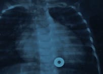
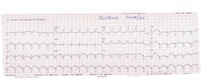
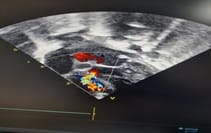
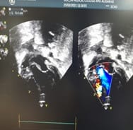
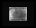
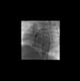
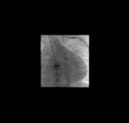
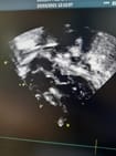
Surgical closure of the ventricular septal defect is a time tested and well-accepted procedure to date especially in smaller babies with failure to thrive and severe pulmonary hypertension. Surgical closure is regarded as the gold standard treatment. However over the past 10 years percutaneous trans-catheter device closure has emerged as a safer alternative especially in the case of muscular VSD. Transcatheter closure of ventricular septal defects (VSD) has not yet received general acceptance. It is not well-established in the younger age group with a weight category of less than 5kg. Occasionally, a hybrid procedure has been proposed. We believe that certain types of VSD can be closed safely by the transcatheter approach even if weight is less than 5kg, especially if the trend of miniaturizing devices continues. This approach should be considered as a valid alternative to a surgical option. We are reporting such a VSD baby with a weight of 2.3 Kg at the age of 6 months, which was closed with a duct occluder.

Keywords: Failure to thrive, Large VSD, Severe PAH, Transcatheter closure

Corresponding Author How to Cite this Article To Browse
Mirza Mohd Kamran, Assistant professor, Department of Pediatric Cardiology, PCE CS unit, JNMCH, AMU, Aligarh, Uttar Pradesh, India.
Email:
Kamran M, Abqari S, Haseen A, Yadav M. Closure of a large upper muscular ventricular septal defect in a 2.3 kg baby using a duct occluder. Pediatric Rev Int J Pediatr Res. 2021;8(3):163-167.
Available From
https://pediatrics.medresearch.in/index.php/ijpr/article/view/670

Manuscript Received Review Round 1 Review Round 2 Review Round 3 Accepted
2021-05-01 2021-05-03 2021-05-15 2021-05-25 2021-06-14
Conflict of Interest Funding Ethical Approval Plagiarism X-checker Note
No Nil Yes 6%

© 2021 by Mirza Mohd Kamran, Shaad Abqari, Azam Haseen, Mayank Yadav and Published by Siddharth Health Research and Social Welfare Society. This is an Open Access article licensed under a Creative Commons Attribution 4.0 International License https://creativecommons.org/licenses/by/4.0/ unported [CC BY 4.0].

Introduction

For decades, surgical closure of ventricular septal defects is a time tested well accepted and also very much acknowledged modality of treatment. Surgical closure of VSD was first portrayed by Lillehei et al. in 1954 and it kept on being viewed as the gold standard treatment [1]. Anyway in recent years percutaneous trans-catheter device closure has emerged as a safer option particularly with muscular VSD. We are reporting an infant, whose weight was 2.3 Kg at 6 months, having an upper muscular VSD which was closed percutaneously with a duct occluder (ADOII).

Clinical summary Our patient is a 6 months old boy who had frank features of congestive heart failure (CHF) and inability to thrive. His weight was 2.3 kg (less than the fifth percentile) and his stature was 52 cm. He looks emaciated and in distress (Fig 1). He was a low birth weight baby with a birth weight of 2.1 Kg, has been diagnosed as a large upper muscular VSD with severe PAH on the 15thday of life. He was put on medical treatment in form of furosemide and enalapril.

On maximum dose of these drugs he couldn’t show any clinical improvement, his congestive heart failure never improved and got admitted multiple times for lower respiratory chest infections (LRTI) including one life-threatening pneumonia for which he was mechanically ventilated 6 days. On assessment, there was tachycardia (HR180/minute), tachypnoea (RR 60/minutes) and the liver was 4 cm beneath the right costal edge. On auscultation first heart sound (S1) was normal but the second heart sound (S2) was loud. There was a grade 2/6 systolic murmur over the left precordium.

Investigations Chest X-Ray showed cardiomegaly and plethoric lung field (Fig2). The electrocardiogram (ECG) showed normal sinus rhythm with superior axis deviation and right ventricular forces (Fig 3). Echocardiography revealed situs solitus, levocardia, intact atrial septum, Mild tricuspid regurgitation (TR) with Gradient of 55 mm HG with moderate Mitral Regurgitation (MR). There was a large upper muscular VSD (Fig 3) with the bidirectional flow (predominantly left-to-right shunt) The VSD estimated 4.5 mm from both right and left ventricle. There was evidence of volume overload in form dilation of the left atrium (LA), Left ventricle (LV) with a left atrial to the aortic ratio (LA/Ao) of 1.8. LV was dilated with and LV end-diastolic dimension

(LVEDD) z-score of more than +3.8. Rest of finding was normal, final impression was large upper muscular VSD with severe pulmonary hypertension (PAH) with evidence of LA and LV dilation.

Procedure At first surgical closure was offered to the patient given refractory congestive heart failure (CHF) and positive history of recurrent lower tract infection (LRTI), out of which the last admission was life-threatening for which the patient was intubated and kept ventilated for six days. With this background, the case was discussed in detail with the cardiovascular surgical team and parents.

The most prominent concerns to go for surgical closure of VSD with this patient was low weight and the need for prolonged postoperative care. Finally, after considering all these issues, it was decided to go for device closure without giving general anesthesia(GA) and also avoiding intubation for obvious reasons. More importantly, encouraging fact with this patient was the location of VSD (upper muscular, distant from tricuspid and aortic valve) and the accessibility of low profile and soft hardware urged us to endeavour VSD device closure with the expectation of utilizing the Amplatzer duct occluder II 6/4 (Abbott, USA).

The technique was performed under conscious sedation (intentionally planned)with a full backup of intubation and provision of general anesthesia (GA). After acquiring femoral venous access (5F) and femoral arterial access (5F), a hemodynamic assessment was first performed to get pressures and saturations.

pedia_670_01.jpg


Figure 1. 6 months old with 2.3 Kg

The pulmonary arterial pressure was 58/19 mmHg with a mean of 38 mmHg against the systemic pressure was 68/24 mmHg with a mean of 43 mmHg (severe PAH). The ratio of pulmonary to systemic blood flow (Qp: Qs) was 3:1 on room air. Since the anatomy of VSD was delineated on Transthoracic echocardiography (TTE) and the Transthoracic view were very good it was decided to avoid LV angiogram to limit the duration of the procedure and also to avoid dye related complication in the already compromised infant. The VSD was crossed with a 5F JR catheter & 0.032 X 260 cm J tipped Terumo wire from LV. The wire was placed in the LPA, 5F guiding JR was taken into RV over the crossed wire.

pedia_670_02.jpg

Figure 2 X-Ray showing significant cardiomegaly (CTR-0.7) with pulmonary plethora

pedia_670_03.jpg

Figure-3: ECG showing the normal sinus rhythm, superior axis, and right ventricular forces

pedia_670_05.jpg

pedia_670_04.jpg

Figure 4 Echo images depicting (a) significant shunt across the ventricular septal defect, grossly dilated left atrium and left ventricle (b)

pedia_670_06.jpg

Figure 5. Showing crossed VSD from LV side, crossed wire is parked in deep LPA

The 6X 4 mm Amplatzer Duct Occluder (II) was deployed under fluoroscopic and transthoracic echocardiography guidance. Finally the device was released which took its predestined shape.

Post-procedure TTE showed the device to be perfectly in place and there was no residual flow. Formation of AV loop and introduction of the long sheath was avoided with this technique. (Fig 6). Total procedure time was around 25 min and fluoroscopy time was 10 min.

Figure 6 Deployment of device


pedia_670_07.jpg pedia_670_08.jpg

Post-procedure the child remained hemodynamically stable; no evidence of hemolysis, ECG showed normal sinus rhythm. Post-intervention echo showed the device an excellent position (Fig 7)

pedia_670_09.jpg

Figure. 7 showing excellent device position

Follow up

After fourteen days, the child began to put on weight (2.8 kg). Only in 20 days all CHF medication was stopped, there was improved oral intake, his symptoms( fast breathing ) was resolved in few days. On follow up Echo there was no residual shunt, no AR, TR and no pericardial effusion. however long term follows up is required.

Discussion

Transcatheter closure of VSD was described in the last part of the 80s and mid-90s [2–3]. While the closure of muscular VSD discovered acknowledgement, it was the perimembranous VSD, which came into dishonour, with past investigations tracking down a high pace of complete atrioventricular block associated with this method [4]. By and large transcatheter closure has been restricted to body weight over 10 kg [5]. Our patient had a bodyweight of 2.3 kg, and for him surgical closure was the first-line treatment option at any given point in time.

The realities were that the patient could not thrive and also he was admitted twice to the emergency unit with congestive heart failure and LRTI which makes him not a decent candidate to go through CPB (cardiopulmonary bypass) which was needed for surgical closure. This urged us to opt for a less invasive option and also the location of VSD (upper muscular, away from TV, AV) made the danger of heart block with transcatheter approach far-fetched because any device placed will be away from the conduction tissue [6].

Off-label utilization of Amplatzer ductal occluders in Perimembranous VSD has been accounted for [7]. As the size of VSD was 4.5 mm, we chose a 6×4 mm duct occluder (ADO II). If anatomy (size and location) of defect can be nicely established on echo, it’s always better to avoid LV angiogram in such small babies as it would reduce the total duration of the procedure and also avoid the injection of contrast agent into the body.

AV loop formation is not free from risk as it stents the small heart like this patient, it’s always better to avoid forming an AV loop and taking larger, longer sheath from the venous side, rather its safer to deploy the device from arterial side (retrograde)in these small babies.


Conclusions

We believe that in such muscular VSD, in babies with low body weight also associated with numerous other risk factors, a transcatheter approach with its low profile hardware and devices may be a decent choice to avoid surgery and its complications in these small kids.

Reference

01. Lillehei Cw, Cohen M, Warden He, Ziegler Nr, Varco Rl. The results of direct vision closure of ventricular septal defects in eight patients by means of controlled cross circulation. Surg Gynecol Obstet. 1955 Oct;101(4)446-66. [Crossref][PubMed][Google Scholar]

02. Chungsomprasong P, Durongpisitkul K, Vijarnsorn C, Soongswang J, Lê TP. The results of transcatheter closure of VSD using Amplatzer® device and Nit Occlud® Lê coil. Catheter Cardiovasc Interv. 2011 Dec 1;78(7)1032-40. doi: 10.1002/ccd.23084 [Crossref][PubMed][Google Scholar]

03. Carminati M, Butera G, Chessa M, De Giovanni J, Fisher G, Gewillig M, et al. Transcatheter closure of congenital ventricular septal defects- results of the European Registry. Eur Heart J. 2007 Oct;28(19)2361-8. doi: 10.1093/eurheartj/ehm314 [Crossref][PubMed][Google Scholar]

04. Yang J, Yang L, Wan Y, Zuo J, Zhang J, Chen W, et al. Transcatheter device closure of perimembranous ventricular septal defects- mid-term outcomes. Eur Heart J. 2010 Sep;31(18)2238-45. doi: 10.1093/eurheartj/ehq240 [Crossref][PubMed][Google Scholar]

05. Moodie DS. Technology Insight- transcatheter closure of ventricular septal defects. Nat Clin Pract Cardiovasc Med. 2005 Nov;2(11)592-6. doi: 10.1038/ncpcardio0351 [Crossref][PubMed][Google Scholar]

06. El Said HG, Bratincsak A, Gordon BM, Moore JW. Closure of perimembranous ventricular septal defects with aneurysmal tissue using the Amplazter Duct Occluder I- lessons learned and medium term follow up. Catheter Cardiovasc Interv. 2012 Nov 15;80(6)895-903. doi: 10.1002/ccd.23074 [Crossref][PubMed][Google Scholar]

07. Dilawar M, Numan M, El-Sisi A, Gendi SM, Ahmad Z. Percutaneous closure of ventricular septal defect associated with tunnel-shaped aneurysm using the Amplatzer duct occluder. Pediatr Cardiol. 2008 Mar;29(2)366-70. doi: 10.1007/s00246-007-9092-0 [Crossref][PubMed][Google Scholar]