E-ISSN:2349-3267
P-ISSN:2349-5499

Research Article

Anterior

Pediatric Review - International Journal of Pediatric Research

2021 Volume 8 Number 4 July-August
Publisherwww.medresearch.in

A Case of Anterior Mediastinal Mass treated as Bronchial Asthma.

Jadhav R.1, Motwani P.2*, Agarkhedkar S.3
DOI: https://doi.org/10.17511/ijpr.2021.i04.07

1 Renuka Jadhav, Professor, Department of Pediatrics, Dr D Y Patil Medical College, Pune, Maharastra, India.

2* Pooja Motwani, Resident, Department of pediatrics, Dr D Y Patil Medical College, Pune, Maharashtra, India.

3 Sharad Agarkhedkar, Head of the Department, Department of pediatrics, Dr D Y Patil Medical College, Pune, Maharashtra, India.

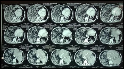
Anterior Mediastinal Masses (AMM) can range in presentation from an asymptomatic incidental finding to significant cardiorespiratory compromise. Symptoms do not always correlate to the degree of compression. Patients with AMM are at high risk for hemodynamic instability and cardiovascular arrest with induction of anesthesia. This report describes a child with a large AMM who presented with classic symptoms of recurrent wheezing and was inadequately evaluated. It is imperative, especially in these unexpected presentations, to have a high index of suspicion.

Keywords: Anterior Mediastinal Mass, Recurrent wheezing, Thymic Lymphoma

Corresponding Author How to Cite this Article To Browse
Pooja Motwani, Resident, Department of pediatrics, Dr D Y Patil Medical College, Pune, Maharashtra, India.
Email:
Renuka Jadhav, Pooja Motwani, Sharad Agarkhedkar, A Case of Anterior Mediastinal Mass treated as Bronchial Asthma.. Pediatric Rev Int J Pediatr Res. 2021;8(4):207-211.
Available From
https://pediatrics.medresearch.in/index.php/ijpr/article/view/680

Manuscript Received Review Round 1 Review Round 2 Review Round 3 Accepted
2021-06-16 2021-06-26 2021-07-10 2021-07-20 2021-07-29
Conflict of Interest Funding Ethical Approval Plagiarism X-checker Note
No Nil Yes 7%

© 2021 by Renuka Jadhav, Pooja Motwani, Sharad Agarkhedkar and Published by Siddharth Health Research and Social Welfare Society. This is an Open Access article licensed under a Creative Commons Attribution 4.0 International License https://creativecommons.org/licenses/by/4.0/ unported [CC BY 4.0].

Introduction

The mediastinum is in the central part of the thoracic cavity and contains many vital organs, such as the heart, trachea, thymus, esophagus, etc. The structure of the mediastinum can be divided into three compartments, namely, the anterior, middle, and posterior mediastinum, from which various types of tumours may originate. [1,2]. Tumours arising from the mediastinum are rare in children and adolescents, and most such tumours are malignant.

For instance, thymomas and lymphomas can arise from the anterior mediastinum. Germ cell tumors mainly arise from the middle mediastinum, while neurogenic tumors often originate from the sympathetic nerve chains or spinal roots of the posterior mediastinum [3]. In children, Lymphoma is the most common primary anterior mediastinal tumor. Germ cell neoplasms are the second most prevalent tumor, followed by thymic lesions and mesenchymal tumors. In children, thymic abnormalities consist primarily of hyperplasia and cysts.

Case report

A 10-year-old boy second issue of non-consanguineous marriage presented to the casualty with complaints of difficulty in breathing and altered mentation for the past couple of hours.

History of similar complaints four months back. He was diagnosed with bronchial asthma with lower respiratory tract infection and was treated for the same. The child had minimal relief with the ongoing treatment—no history of bronchial asthma or allergies in the family. On examination child was toxic looking, GCS 8/15, with features of hypovolemic shock, had severe pallor and pedal edema. On cardiovascular examination, the patient had gallop rhythm. On respiratory system examination trachea was shifted to the left side and had a silent chest on auscultation.

The child was admitted to PICU, and attempts were made for hemodynamic stabilization was child put on mechanical ventilator support. Within 48 hours of stay in PICU child had multiple fever spikes, one episode of Generalized Tonic-clonic convulsion for which adequate treatment was started. The child had a bout of desaturation when high-quality CPR was started but could not be revived.

Investigations-The chest X-ray film showed opacification of the right upper- and mid-Hemithorax. Computed tomography (CT) showed a sizeable mixed density soft tissue lesion almost entirely occupying the anterior mediastinum, prevascular space and almost entire right hemithorax with resultant compression atelectasis of the right lung. Hemidiaphragm was compressed inferiorly, and there was a cardiomediastinal shift to the left due to lesion. Blood reports were suggestive of microcytic hypochromic anemia, leukocytosis, thrombocytopenia, deranged liver and renal function test and abnormal coagulation profile. ABG was suggestive of severe uncompensated metabolic acidosis.

2D-echo suggestive of global LV hypokinesia with EF of 25%. Sonogram of abdomen and pelvis suggestive of normal study. Image-guided lung biopsy was done under RDP cover. Biopsy showed the presence of mature lymphocytes, hassells corpuscles like structure, lymphoid cells, mature adipose tissue and fibro collagenous tissue. No evidence of sarcomatous elements. Based on anterior mediastinal mass with thymic tissue, possibilities considered were thymic mass hyperplasia or thymic neoplasm.

pedia_680_01.jpg

Figure 1: shows a large mixed density soft tissue lesion almost entirely occupying the anterior mediastinum, prevascular space and almost entire right hemithorax with resultant compression atelectasis of the right lung.

Discussion

The thymus, which is an anterior mediastinal organ, weighs approximately 25 g during birth. It reaches a maximum weight of 30–40 g during puberty and then undergoes involution but not disappear completely. However, its percentage ratio to body weight is maximum at birth (0.76) and reaches 0.19 at around five years of age [4].


Even the shape of the thymus changes with age where it is known to be quadrilateral shaped with convex margins at around five years of age and changes to triangular shape with straight margins by the age of 15 years [5]. Thymic lesions form approximately 2% of all mediastinal tumours in children [6]. The clinical profile of these patients varies from asymptomatic incidentally detected masses to being symptomatic with dyspnoea, dysphagia or venous congestion [8]. The myriad paraneoplastic syndromes (PNS) associated with the thymus, such as myasthenia gravis (MG), pure red cell aplasia, hypogammaglobulinemia, pemphigus, Sjogren's syndrome, manifest more commonly in adults rather than in childhood [1,7]. Besides the ones mentioned above, other clinical manifestations include gastrointestinal disorders (chronic ulcerative colitis), collagen and autoimmune disorders (scleroderma and polymyositis), dermatologic disorders (alopecia), endocrine disorders (Cushing's syndrome), renal disease (nephrosis) and hematologic syndromes (agranulocytosis) [7]. It has also been seen that MG is the most common PNS and is generally seen commonly in children <10 years of age [8].

Radiological classification Radiologists have attempted to classify thymic lesions in children according to the density of the visible mass into solid, fatty and cystic lesions. An ordinary differential of a solid thymic mass in a child is a prominent normal thymus which is also referred to as a "pseudomass" since it isn't a tumor but the normal appearance of the thymus in that particular age group [5]. The other solid lesions are ectopic thymus, retrocaval (between the superior vena cava and the great arteries) or cervical, thymic hyperplasia or thymic neoplasms. The cystic lesions include thymic cysts, and fatty lesions are the thymolipomas.

The other method by which radiologists have classified paediatric thymic lesions is based on the size of the thymus, whether it is small or large [9]. Small thymus can be physiological, which may be age-related involutions or treatment-related atrophy or immunodeficiency disorders leading to hypoplasia or aplasia. The large thymus included hyperplasias and neoplastic lesions. Thymic hyperplasia can be true thymic hyperplasia or lymphoid (follicular) hyperplasia. True thymic hyperplasias result in a change in the shape of the gland and an increase in the size, which may be more than 50% of the original.

The hyperplasia is seen in patients recovering from various stressors such as burns, significant surgeries, infections, post therapies which may be corticosteroids, radiation or chemotherapy, or they may have other related disorders such as hyperthyroidism or sarcoidosis. Thymic hyperplasia may present as an emergency with symptoms of respiratory stridor, dysphagia, cyanosis and dyspnea. [10].

Thymic cysts are uncommon among the paediatric population forming less than 1% of all mediastinal masses and may be congenital or acquired. The congenital cysts also arise along the thymopharyngeal duct when the thymus undergoes atrophy, and the remnants undergo cystic change [11]. They present suddenly as painless neck masses and are commoner on the left side of the neck, more prevalent on the anterior surface of sternocleidomastoid, and around 50% of them may extend to the superior mediastinum [12,13]. The acquired cysts may arise post-radiotherapy in the case of Hodgkin or non-Hodgkin Lymphoma or as inflammatory cysts seen in autoimmune disorders. They may also occur post-thoracotomy or may be associated with thymic tumours that cause distortion and compression of surrounding normal thymic tissue [14]. The congenital cysts are usually unilocular, while those arising in the thymus are multilocular, occurring due to degeneration of Hassal's corpuscles [13].  A significant differential of thymic cysts is a cystic thymoma which is challenging to distinguish radiologically. The histopathological features clinch the diagnosis and include the absence of epithelial lining of the cyst wall, solid areas in the wall comprising of epithelial cells and lymphoid cells and perivascular spaces along with medullary differentiation at some foci [15].

Thyomyolipomas are benign neoplasms comprising of normal thymic tissue interspersed with fat and fibrous septae. Due to their fat content, they are incredibly pliable and may sometimes occupy the entire thoracic cavity [14]. They may be incidentally detected, or the patients may present with compression symptoms or associated PNS. They have encapsulated non-infiltrating lesions located mainly in the inferior part of the anterior mediastinum. Its diagnosis can be proposed radiologically due to its appearance of a fatty mass, location and continuity with the thymus. Thymic epithelial neoplasm includes thymoma, thymic carcinoma, and thymic carcinoid.


Thymomas are extremely rare in children forming less than 5% of all mediastinal masses. They are usually asymptomatic and are detected incidentally on routine chest radiography. Thymic carcinomas are sporadic tumors in adults and are even rarer in the paediatric age group forming less than 1% of all childhood neoplasms [16,17]. The thymic carcinomas arise from the thymic epithelial cells, which lose their functional and phenotypic features and appear anaplastic [6]. Thymic carcinoids are also rare, with an incidence of 2.6–8% [18]. They are exclusively found in males. These carcinoids are known to have increased morbidity with local spread, distant metastasis and recurrence.

Thymic Lymphoma forms the most common anterior mediastinal mass in children, and thymic involvement by lymphomas can occur commonly in disseminated disease compared to primary thymic origin. Among the histopathological subtypes, Hodgkin lymphoma forms the most common variant [14].

Conclusion

This case shows the importance of evaluating any infant or child presenting with recurrent wheezing for anterior mediastinal mass. Suspicion on initial presentation is a must as the prognosis and survival rates are highly dependent on early diagnosis and treatment.

Reference

01. Thacker PG, Mahani MG, Heider A, Lee EY. Imaging Evaluation of Mediastinal Masses in Children and Adults: Practical Diagnostic Approach Based on A New Classification System. J Thorac Imaging. 2015 Jul;30(4):247-67. doi: 10.1097/RTI.0000000000000161 [Crossref][PubMed][Google Scholar]

02. McCarville MB. Malignant pulmonary and mediastinal tumors in children: differential diagnoses. Cancer Imaging. 2010 Oct 4;10 Spec no A(1A):S35-41. doi: 10.1102/1470-7330.2010.9015 [Crossref][PubMed][Google Scholar]

03. Takeda S, Miyoshi S, Akashi A, Ohta M, Minami M, Okumura M, et al. Clinical spectrum of primary mediastinal tumors: a comparison of adult and pediatric populations at a single Japanese institution. J Surg Oncol. 2003 May;83(1):24-30. doi: 10.1002/jso.10231 [Crossref][PubMed][Google Scholar]

04. Ocal T, Türken A, Ciftçi AO, Senocak ME, Tanyel FC, Büyükpamukçu N. Thymic enlargement in childhood. Turk J Pediatr. 2000 Oct-Dec;42(4):298-303. [Crossref][PubMed][Google Scholar]

05. Ranganath SH, Lee EY, Restrepo R, Eisenberg RL. Mediastinal masses in children. AJR Am J Roentgenol. 2012 Mar;198(3):W197-216. doi: 10.2214/AJR.11.7027 [Crossref][PubMed][Google Scholar]

06. Yaris N, Nas Y, Cobanoglu U, Yavuz MN. Thymic carcinoma in children. Pediatr Blood Cancer. 2006 Aug;47(2):224-7. doi: 10.1002/pbc.20468 [Crossref][PubMed][Google Scholar]

07. Stachowicz-Stencel T, Orbach D, Brecht I, Schneider D, Bien E, Synakiewicz A, et al. Thymoma and thymic carcinoma in children and adolescents: a report from the European Cooperative Study Group for Pediatric Rare Tumors (EXPeRT). Eur J Cancer. 2015 Nov;51(16):2444-52. doi: 10.1016/j.ejca. 2015.06.121 [Crossref][PubMed][Google Scholar]

08. Saha S, Suhani S, Basak A, Agarwal N, Dewan P. Pediatric thymoma with a difference: report of a case and review of literature. J Surg Tech Case Rep. 2014 Jul-Dec;6(2):64-6. doi: 10.4103/2006-8808.147263 [Crossref][PubMed][Google Scholar]

09. Manchanda S, Bhalla AS, Jana M, Gupta AK. Imaging of the pediatric thymus: Clinicoradiologic approach. World J Clin Pediatr. 2017 Feb 8;6(1):10-23. doi: 10.5409/wjcp.v6.i1.10 [Crossref][PubMed][Google Scholar]

10. Mitchell a Graeme, and josef warkany. "The problem of the thymus in children". Journal of the American Medical Association. 112;4(1939): 283-285. [Crossref][PubMed][Google Scholar]

11. Nasseri F, Eftekhari F. Clinical and radiologic review of the normal and abnormal thymus: pearls and pitfalls. Radiographics. 2010 Mar;30(2):413-28. doi: 10.1148/rg.302095131 [Crossref][PubMed][Google Scholar]

12. Prosser JD, Myer CM 3rd. Branchial cleft anomalies and thymic cysts. Otolaryngol Clin North Am. 2015 Feb;48(1):1-14. doi: 10.1016/j.otc. 2014.09.002 [Crossref][PubMed][Google Scholar]

13. Sturm JJ, Dedhia K, Chi DH. Diagnosis and Management of Cervical Thymic Cysts in Children. Cureus. 2017 Jan 11;9(1):e973. doi: 10.7759/cureus.973 [Crossref][PubMed][Google Scholar]


14. Goldstein AJ, Oliva I, Honarpisheh H, Rubinowitz A. A tour of the thymus: a review of thymic lesions with radiologic and pathologic correlation. Can Assoc Radiol J. 2015 Feb;66(1):5-15. doi: 10.1016/j.carj.2013.09.003 [Crossref][PubMed][Google Scholar]

15. Honda S, Morikawa T, Sasaki F, Okada T, Naito S, Itoh T, et al. Cystic thymoma in a child: a rare case and review of the literature. Pediatr Surg Int. 2007 Oct;23(10):1015-7. doi: 10.1007/s00383-007-1988-5 [Crossref][PubMed][Google Scholar]

16. Sekihara K, Okuma Y, Kawamoto H, Hosomi Y. Clinical outcome of thymic lymphoepithelioma-like carcinoma: Case report of a 14-year-old male. Oncol Lett. 2014 Nov;8(5):2183-2186. doi: 10.3892/ol.2014.2475 [Crossref][PubMed][Google Scholar]

17. Ferrari A, Bisogno G, De Salvo GL, Indolfi P, Perilongo G, Cecchetto G; Italian Study on Rare Tumours in Paediatric Age (TREP); Associazione Italiana Ematologia Oncologia Pediatrica (AIEOP). The challenge of very rare tumours in childhood: the Italian TREP project. Eur J Cancer. 2007 Mar;43(4):654-9. doi: 10.1016/j.ejca.2006.08.028 [Crossref][PubMed][Google Scholar]

18. Singh Ospina N, Thompson GB, C Nichols F 3rd, Cassivi SD, Young WF Jr. Thymic and Bronchial Carcinoid Tumors in Multiple Endocrine Neoplasia Type 1: The Mayo Clinic Experience from 1977 to 2013. Horm Cancer. 2015 Dec;6(5-6):247-53. doi: 10.1007/s12672-015-0228-z [Crossref][PubMed][Google Scholar]