E-ISSN:2349-3267
P-ISSN:2349-5499

Case Report

Solitary Parotid Cysticercosis

Pediatric Review - International Journal of Pediatric Research

2022 Volume 9 Number 5 September October
Publisherwww.medresearch.in

Solitary parotid cysticercosis in a 7-year-old boy: A rare entity

Katyayani K M.1*, Devi K M.2, Sreevalli Sarma S S.3, Krishna Reddy K. V.4, Krishna P V.5
DOI: https://doi.org/10.17511/ijpr.2022.i05.04

1* Mounika Katyayani K, Consultant, Pediatrics, Community health center, Narsipatnam, Andhra Pradesh, India.

2 Mamatha Devi K, Department of Otorhinolaryngology, Maheshwara Medical College, Hyderabad, Telangana, India.

3 Sai Sreevalli Sarma S, Department of Community Medicine, Katuri Medical College, Guntur, Andhra Pradesh, India.

4 Vamsi Krishna Reddy K., Department of Pediatrics, Indira medical college, Thiruvallur, Tamil Nadu, India.

5 Vamsi Krishna P, NRI medical college, Guntur, Andhra Pradesh, India.

Cysticercosis is a systemic parasitic illness caused by larval forms of Tinea solium. Being acquired by feco-oral contamination, cysticercosis commonly affects the central nervous, skeletal, cardiac, skin, and subcutaneous tissues. However, isolated parotid gland cysticercosis is extremely rare. In this report, the authors present a rare case of a 7-year-old boy who presented with left-sided parotid swelling, diagnosed as cysticercosis by imaging and fine needle aspiration cytology. The boy was managed by both surgical and medical modalities and was put under regular follow-up.

Keywords: Cysticercosis, Tinea solium, parotid swelling, fine needle aspiration cytology

Corresponding Author How to Cite this Article To Browse
Mounika Katyayani K, Consultant, Pediatrics, Community health center, Narsipatnam, Andhra Pradesh, India.
Email:
Mounika Katyayani K, Mamatha Devi K, Sai Sreevalli Sarma S, Vamsi Krishna Reddy K., Vamsi Krishna P, Solitary parotid cysticercosis in a 7-year-old boy: A rare entity. Pediatric Rev Int J Pediatr Res. 2022;9(5):51-54.
Available From
https://pediatrics.medresearch.in/index.php/ijpr/article/view/730

Manuscript Received Review Round 1 Review Round 2 Review Round 3 Accepted
2022-10-08 2022-10-10 2022-10-17 2022-10-24 2022-10-31
Conflict of Interest Funding Ethical Approval Plagiarism X-checker Note
Nil Nil Yes 17%

© 2022by Mounika Katyayani K, Mamatha Devi K, Sai Sreevalli Sarma S, Vamsi Krishna Reddy K., Vamsi Krishna Pand Published by Siddharth Health Research and Social Welfare Society. This is an Open Access article licensed under a Creative Commons Attribution 4.0 International License https://creativecommons.org/licenses/by/4.0/ unported [CC BY 4.0].

Introduction

Cysticercosis is a systemic parasitic illness caused by larval forms of Tinea solium. Intense transmission of this cestode occurs in central and south America, Southeast Asia, and Sub-Saharan Africa, where poor sanitation facilities exist, resulting in feco-oral contamination of food and water [1].

Travelling to these endemic areas has an increased risk of acquiring the disease.

Cysticercosis commonly affects the central nervous system, skeletal and cardiac muscle, skin, and subcutaneous tissues. Isolated parotid gland involvement is extremely rare [2,3].

The disease induces symptoms predominantly due to dead and degenerating cysticercal cysts.

Serology, imaging, fine needle aspiration cytology (FNAC), and routine hematological and stool workup could aid in diagnosis.

Histopathological examination of aspirate would reveal cysticercal forms that directed towards therapeutic intervention. The author hereby present a case of a 7-year-old boy with parotid gland cysticercosis for its rarity.

Case Presentation

A 7-year-old boy, a student of second grade, was accompanied by his mother to the pediatric clinic with a chief complaint of swelling in front of his left ear for the past 3months.

The mother initially noted a small swelling followed by a gradual increase over the course. The child had no history of pain, fever, trauma, weight loss, easy fatiguability, bleeding manifestations or bone pains. There was no significant past history.

The child was born to a non-consanguineous couple and was second in birth order. He belonged to a low socio-economic class. He resides in a pucca house, in a rural locality in Guntur, India with several lakes close to his vicinity.

His growth and development were normal. He consumes a variety of plant and animal-based foods; however, with no significant deficits in calories and protein. There was no significant family history. The boy was immunized as per the national immunization schedule.

Upon general examination, the child was well nourished, conscious, coherent, and oriented to time, place, and person.

There were no dysmorphic facies, neurocutaneous or bleeding markers. Limbs and posture were normal. Vitals were stable. The child’s anthropometry was normal.

Upon local examination, a single, ovoid swelling was present in the left pre-auricular region (Figure-1), measuring 3cms×2cms, with regular borders and soft consistency.

It was mobile, non-tender, non-pulsatile, and non-compressible, with normal skin.

pedia_730_01.JPG

Figure 1: left pre-auricular swelling in a 7-year-old boy (pointed by an arrow)

Upon neck examination, a single lymph node was palpable at the left upper jugular area, measured 2cms×2cms.

It was mobile, firm, and non-tender, with intact skin.

Systemic examination revealed no abnormalities.

By clinical presentation, a differential diagnosis of Acute infectious parotitis, preauricular lymphadenitis, lipoma, benign epithelial tumors, and vascular malformations have been considered.

An extensive diagnostic workup included a basic hemogram, with an elevated eosinophil count and erythrocyte sedimentation rate, normal electrolyte panel. Ultrasound features of the lesion were shown (Figure 2).


pedia_730_02.JPGFigure 2: USG of the lesion showing 2.3cm×1.4cm hypoechoic cystic lesion with central calcification focus (marked by arrow) over the left lower lobe of the parotid

FNAC of the parotid lesion yielded straw-colored fluid. Air-dried smears from aspirate, revealed a suspicious structure with degenerated scolex, neck and proglottids in congruence with a cestode infection (Figure 3).

pedia_730_03.JPG
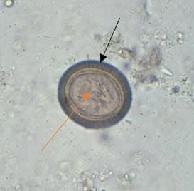
Figure 3: Histopathological examination (H & E,100x) of aspirate showing structure with scolex (black arrow), suckers and hooks (green arrow), neck (blue arrow) and proglottid (red arrow)

in an inflammatory background. However, serology revealed positive anticysticercal antibodies and stool examination showed Tinea solium egg (Figure-4) clinching a definitive diagnosis.

pedia_730_04.JPG

Figure 4: Stool microscopy (40x) showing Tinea solium egg with a thick radiate shell (black arrow) and hooklets (red arrow).

FNAC of the left upper jugular lymph node showed inflammatory infiltration with no neoplastic cells. Anti-helminthic therapy was initiated with albendazole at 15mg/kg/day orally in two divided doses for 7days. After a month of follow-up, the child showed significant improvement.

Discussion

Tinea solium (pork tapeworm), is the second most common cestode infection in humans [3]. It has a scolex with suckers and hooklets, a neck and numerous proglottids with both male and female reproductive structures [2]. In its complex life cycle, the pig acts as an intermediate and the man as a definitive host [1,2].

Children acquire the cestode by ingestion of undercooked pork containing larval cysts and from the eggs shed by tapeworm carriers. After ingestion, adult tapeworm form is attained in the intestine, which migrates to the bloodstream and to various tissues, where they form cysticercal cysts. Most cysts remain asymptomatic for a few years; symptoms typically manifest when they begin to degenerate, associated with a host inflammatory response.


Clinical presentation depends on cysticercal location. Cantey et al reported 2% of emergency department visits due to seizures were due to neurocysticercosis [1]. However, cysticercal presence in oral mucosa and parotid gland is extremely rare. While Sharma and Kaur reported only 38 cases of oral mucosal cysticercosis in their research, only 5 cases of parotid cysticercosis have been reported so far in the literature up to 2017 [4,5,6,7]. The current report uplifts consideration of cysticercosis in a case of isolated parotid mass.

Epidemiological exposure, clinical presentation, sonographic findings, and histopathological analysis serve as the main diagnostic tool for detecting cysticercosis. While typical USG findings demonstrated hypoechoic cystic lesions [5], FNAC shows inflammatory infiltration with a granular background [7]. The FNAC success rate allowed physicians to early initiation of anti-helminthic therapy and thereby prevent disseminated disease course. Nevertheless, Veena et al [8] delineated the need for surgery along with medical therapy in certain cases.

Conclusion

This rare entity of solitary parotid mass in a 7year old boy spotlights diagnostic consideration of cysticercosis, especially in patients residing in endemic areas.

Reference

01. Cantey PT, Coyle CM, Sorvillo FJ, Wilkins PP, Starr MC, Nash TE. Neglected parasitic infections in the United States: cysticercosis. Am J Trop Med Hyg. 2014 May;90(5):805-809. doi: 10.4269/ajtmh.13-0724 [Crossref][PubMed][Google Scholar]

02. Kumar V, Abbas AK, Fausto N, Robbins SL, Cotran RS, editors. Robbins and Cotran pathologic basis of disease. 7th ed. Philadelphia: Elsevier Saunders; 2005. 1525 p. ISBN: 978-0-7216-0187-8 [Crossref][PubMed][Google Scholar]

03. Carlos R, Contreras E, Rivera H, Gálvez M. Extracranial head and neck cysticercosis: Report of nine cases with emphasis in serologic analysis and natural history of the disease. " Oral Surgery, Oral Medicine, Oral Pathology, Oral Radiology, and Endodontology 98. 2 (2004): 196. [Crossref][PubMed][Google Scholar]

04. P. D. Sharma and M. Kaur, “Cysticercosis of the parotid gland: a case report, International Journal of Medical Research Professionals, vol. 3, no. 3, pp. 291–293, 2017. DOI: [Article][Crossref][PubMed][Google Scholar]

05. Chakraborty PP, Bhattacharjee R, Chatterjee K. Parotid gland cysticercosis. J Assoc Physicians India. 2007 Oct;55:717. [Crossref][PubMed][Google Scholar]

06. Seith A, Gadodia A, Sharma R. Solitary cysticercosis (tapeworm) of the parotid gland. Ear Nose Throat J. 2010 Nov;89(11):522-4. [Crossref][PubMed][Google Scholar]

07. Goyal P, Sehgal S, Ghosh S, Mittal D, Kumar A, Singh S. A cytological study of palpable superficial nodules of parasitic origin: a study of 41 cases. Patholog Res Int. 2014;2014:373472. doi: 10.1155/2014/373472 [Crossref][PubMed][Google Scholar]

08. Veena G, Shon GM, Usha K, Nayar RC. Extracranial cysticercosis of the parotid gland: a case report with a review of the literature. J Laryngol Otol. 2008 Sep;122(9):1008-11. doi: 10.1017/S0022215107000175 [Crossref][PubMed][Google Scholar]