E-ISSN:2349-3267
P-ISSN:2349-5499

Case Report

Ashy dermatosis

Pediatric Review - International Journal of Pediatric Research

2022 Volume 9 Number 5 September-October
Publisherwww.medresearch.in

Ashy Dermatosis – A rare case in a 7year old Indian male child

Sandeep B.1, Shefali P.2*, Kiran B.3, Namitha.4
DOI: https://doi.org/10.17511/ijpr.2022.i05.03

1 B Sandeep, Professor, Department of Pediatrics, East Point College of Medical Sciences & RC, Bangalore, Karnataka, India.

2* P Shefali, Assistant Professor, Department of Pediatrics, East Point College of Medical Sciences & RC, Bangalore, Karnataka, India.

3 B Kiran, Professor & HOD, Department of Pediatrics, East Point College of Medical Sciences & RC, Bangalore, Karnataka, India.

4 Namitha, Assistant Professor, Department of Dermatology, East Point College of Medical Sciences & RC, Bangalore, Karnataka, India.

Ashy dermatosis also known as erythema dyschromicum perstans, is an acquired chronic pigmentary skin disorder. This disorder has been very rarely reported in Indian children. Here we report a case of a 7year old male child presented to pediatrics OPD with multiple areas of hyperpigmentation. A skin biopsy was done which was suggestive of Ashy dermatosis. Early recognition of this condition is important for proper diagnosis and to prevent unnecessary investigations.

Keywords: lichen planus pigmentosus, erythema dyschromicum perstans, ashy dermatosis, skin biopsy, hyperpigmentation

Corresponding Author How to Cite this Article To Browse
P Shefali, Assistant Professor, Department of Pediatrics, East Point College of Medical Sciences & RC, Bangalore, Karnataka, India.
Email:
B Sandeep, P Shefali, B Kiran, Namitha, Ashy Dermatosis – A rare case in a 7year old Indian male child. Pediatric Rev Int J Pediatr Res. 2022;9(5):46-50.
Available From
https://pediatrics.medresearch.in/index.php/ijpr/article/view/732

Manuscript Received Review Round 1 Review Round 2 Review Round 3 Accepted
2022-09-22 2022-09-24 2022-10-01 2022-10-08 2022-10-15
Conflict of Interest Funding Ethical Approval Plagiarism X-checker Note
Nil Nil Yes 18%

© 2022by B Sandeep, P Shefali, B Kiran, Namithaand Published by Siddharth Health Research and Social Welfare Society. This is an Open Access article licensed under a Creative Commons Attribution 4.0 International License https://creativecommons.org/licenses/by/4.0/ unported [CC BY 4.0].

Introduction

Ashy dermatosis or erythema dyschromicum perstans is an unknown entity which occurs worldwide in all races. It is a very rare condition in children, only a few cases have been published as of now in Indian children [1].

It is characterized by oval, polycyclic, irregularly shaped, grey-blue hyperpigmented macules on the trunk, arms, face and neck [2].

The disease has been called ashy dermatosis because of its peculiar slate-like grey discolouration which can present with an erythematous raised border at the beginning stage [3].

The condition was first described in 1957 by Oswaldo Ramirez in salvadorance, who called patients of this condition Los ecenicienta which in Spanish means the ash colored ones because of their characteristic ashy-coloured lesions [4].

The sites commonly involved are the face, neck, trunk and upper limbs but any region of the body can be affected [5].

pedia_1732_01.jpg

Figure 1: bluish grey hyperpigmented lesion visible on left lumbar region of abdomen

pedia_1732_02.JPG

Figure 2: hyperpigmented lesion seen over right deltoid region

pedia_1732_03.JPG
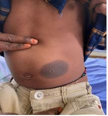
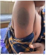
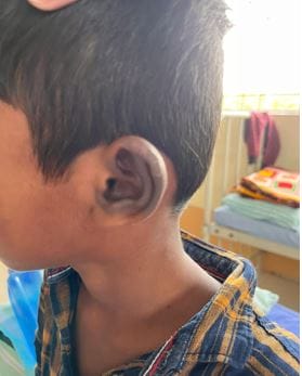
Figure 3: hyperpigmentary changes at the left pinna


pedia_1732_04.jpgFigure 4: hyperpigmented mark with erythematous margin visible over the medial side of left knee joint

pedia_1732_05.jpg
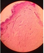
Figure 5: Histopathological examination of biopsy section of the erythematous lesion suggestive of hyperkeratosis of the epidermis with focal vacuolar degeneration of basal layer. The dermis shows increased pigment incontinence and mild perivascular infiltrate.

Case report

A 7-year old dark-skinned male child of Indian origin presented to pediatrics OPD with hyperpigmented flat lesions over the abdomen, right shoulder, left ear and left leg for the last 6moths of duration. Initially, the lesions were pea-sized later enlarged by about 6-7 times. The child did not have any complaints of fever, itching, fatigue or symptoms suggestive of any systemic disease. There was no history of any drug usage before the onset of lesions. On detailed skin examination, the following findings were revealed: brownish grey-coloured, isolated macular lesions over the abdomen at the left lumbar region next to the umbilicus, cylindrical in shape, measuring around 5 ´ 10cm [figure 1]. The second lesion was over the right shoulder at the deltoid region, cylindrical measuring about 6 ´ 12cm [figure 2]. The third lesion over the left ear involves almost 3/4th of the pinna [figure3]. The fourth lesson was found over the left lower limb circular in shape 3 ´ 3cm in diameter [figure 4]. The mucous membrane, palms, soles, nails and back were normal. The systemic examination of this child was normal. All routine investigations were normal, a dermatologist's opinion was taken and a skin biopsy was done which was suggestive of ashy dermatosis [figure 5]. A diagnosis of ashy dermatosis was made based on clinical and histopathological findings.

The child was asked for a follow-up after 3months. The child was seen after 4weeks of treatment and a relative decrease in the size of lesions was noticed then the child was lost to follow-up.

Discussion

Ashy dermatosis is an idiopathic acquired macular hyperpigmentation disorder. The descriptive term ashy or “los cenicientos” was used because of the ashy blue-grey colour of the lesion [6]. Convit, Kerdel-Vegas and Rodriguez in their case series reported the presence of raised erythematous borders in the early stages and proposed the term erythema dyschromicum perstans [7]. Other authors however have noticed that erythema dyschromicum perstans can occur without marginal erythema [8,9]. Bhutani and his colleagues described similar hyperpigmented macules over the flexures and photo-exposed areas


in 40 Indian patients, about a third of whom had associated features of lichen planus on clinical evaluation and histopathological examination [10]. The etiology of ashy dermatosis is unknown. It has been observed that some factors like ingestion of ammonium nitrite, nematodes infestation, contrast media used for x-rays, cobalt toxicity, and exposure to chlorothalonil can also predispose to the development of ashy dermatosis [11,12,13,14]. Ashy dermatosis is slow in onset and is unlikely to resolve spontaneously as compared to children in whom it might resolve within 2-3 years [15]. The treatment modalities for ashy dermatosis include dapsone, clofazimine, antibiotics, steroids, multivitamins, and antihistamines. However, the outcome of these treatment modalities is subjective [16].

Conclusion

Ashy dermatosis to be included in the differential diagnosis of hyperpigmented macules in Indian children. Early detection of this condition will avoid unnecessary investigations and will lead to a relevant diagnosis.

Reference

01. Keisham C, Sarkar R, Garg VK, Chugh S. Ashy dermatosis in an 8-year-old Indian child. Indian Dermatol Online J. 2013 Jan;4(1):30-2. doi: 10.4103/2229-5178.105466 [Crossref][PubMed][Google Scholar]

02. Nieuweboer-Krobotova L. Hyperpigmentation: types, diagnostics and targeted treatment options. J Eur Acad Dermatol Venereol. 2013 Jan;27 Suppl 1:2-4. doi: 10.1111/jdv.12048 [Crossref][PubMed][Google Scholar]

03. Melo CR, Sá MC, Carvalho S. Erythema dyschromicum perstans in a child following an enteroviral meningitis. An Bras Dermatol. 2017 Jan-Feb;92(1):137-138. doi: 10.1590/abd1806-4841.201745144 [Crossref][PubMed][Google Scholar]

04. Leung AKC, Lam JM. Erythema Dyschromicum Perstans in an 8-Year-Old Indian Child. Case Rep Dermatol Med. 2018 Jul 15;2018:2143089. doi: 10.1155/2018/2143089 [Crossref][PubMed][Google Scholar]

05. Cutrì FT, Ruocco E, Pettinato G, Ciancia G.

Lichen planus pigmentosus-like ashy dermatosis. Dermatol Reports. 2011 Dec 6;3(3):e46. doi: 10.4081/dr.2011.e46 [Crossref][PubMed][Google Scholar]

06. Amatya, Bibush. Ashy dermatosis: A comprehensive review. Our Dermatology Online 8. 2 (2017): 143. [Crossref][PubMed][Google Scholar]

07. Leung N, Oliveira M, Selim MA, McKinley-Grant L, Lesesky E. Erythema dyschromicum perstans: A case report and systematic review of histologic presentation and treatment. Int J Womens Dermatol. 2018 Sep 27;4(4):216-222. doi: 10.1016/j.ijwd.2018.08.003 [Crossref][PubMed][Google Scholar]

08. Schwartz RA. Erythema dyschromicum perstans: the continuing enigma of Cinderella or ashy dermatosis. Int J Dermatol. 2004 Mar;43(3):230-2. doi: 10.1111/j.1365-4632.2004.02001.x [Crossref][PubMed][Google Scholar]

09. Muñoz C, Chang AL. A case of Cinderella: erythema dyschromicum perstans (ashy dermatosis or dermatosis cinecienta). Skinmed. 2011 Jan-Feb;9(1):63-4. [Crossref][PubMed][Google Scholar]

10. Bhutani LK, Bedi TR, Pandhi RK, Nayak NC. Lichen planus pigmentosus. Dermatologica. 1974;149(1):43-50. doi: 10.1159/000251470 [Crossref][PubMed][Google Scholar]

11. Jablonska S. Ingestion of ammonium nitrate as a possible cause of erythema dyschromicum perstans (ashy dermatosis). Dermatologica. 1975;150(5):287-91. doi: 10.1159/000251444 [Crossref][PubMed][Google Scholar]

12. Osswald SS, Proffer LH, Sartori CR. Erythema dyschromicum perstans: a case report and review. Cutis. 2001 Jul;68(1):25-8. [Crossref][PubMed][Google Scholar]

13. Zenorola P, Bisceglia M, Lomuto M. Ashy dermatosis associated with cobalt allergy. Contact Dermatitis. 1994 Jul;31(1):53-4. doi: 10.1111/j.1600-0536.1994.tb01911.x [Crossref][PubMed][Google Scholar]

14. Penagos H, Jimenez V, Fallas V, O'Malley M, Maibach HI. Chlorothalonil, a possible cause of erythema dyschromicum perstans (ashy dermatitis). Contact Dermatitis. 1996 Oct;35(4):214-8.


doi: 10.1111/j.1600-0536.1996.tb02360.x [Crossref][PubMed][Google Scholar]

15. Silverberg NB, Herz J, Wagner A, Paller AS. Erythema dyschromicum perstans in prepubertal children. Pediatr Dermatol. 2003 Sep-Oct;20(5):398-403. doi: 10.1046/j.1525-1470.2003.20505.x [Crossref][PubMed][Google Scholar]

16. Torrelo A, Zaballos P, Colmenero I, Mediero IG, de Prada I, Zambrano A. Erythema dyschromicum perstans in children: a report of 14 cases. J Eur Acad Dermatol Venereol. 2005 Jul;19(4):422-6. doi: 10.1111/j.1468-3083.2005.01203.x [Crossref][PubMed][Google Scholar]