E-ISSN:2349-3267
P-ISSN:2349-5499

Case Report

William Syndrome

Pediatric Review - International Journal of Pediatric Research

2023 Volume 10 Number 3 May-June
Publisherwww.medresearch.in

William Syndrome Presenting with Life-threatening Heart failure: Rare case report

Mehrotra A.1*, Shakya U.2, Kacker S.3
DOI: https://doi.org/10.17511/ijpr.2023.i03.05

1* Akhil Mehrotra, Chief, Non Invasive Cardiologist Pediatric and Adult Cardiology, Prakash Heart Station, D-16 Nirala Nagar, Lucknow, Uttar Pradesh, India.

2 Ujala Shakya, Cardiac Technician, Prakash Heart Station, D-16 Nirala Nagar, Lucknow, Uttar Pradesh, India.

3 Shubham Kacker, Lead PMO, Tech Mahindra, New Delhi, India.

Williams syndrome (WS), also referred to as Williams-Beuren syndrome, is a rare complex congenital developmental multisystem disorder, occurring in 1 per 20,000 live births, It is characterized by congenital heart defects (CHD), skeletal, renal anomalies, cognitive disorder, social personality disorder and notably dysmorphic Elfin-like facies. Supravalvular aortic stenosis is the most frequent cardiovascular abnormality in WS children. WS occurs as the result of a deletion of approximately 1.5-1.8 Mb on chromosome 7q11.23. The deletion is almost always denovo, however, familial cases have been reported. A genetic study is usually required for a definitive diagnosis, but genetic testing is often unavailable in developing countries and the combination of a typical clinical phenotype and echocardiographic profile helps to confirm the diagnosis. We are reporting a rare case of WS in a 2-month infant presenting with heart failure because of multiple CHDs, including coarctation of the aorta (COA), patient ductus arteriosus (PDA), and mild supravalvular pulmonary stenosis (SVPS), severe-pulmonary hypertension and systemic hypertension.

Keywords: Heart failure, COA, PDA, Supra-Valvular Pulmonary Stenosis, Elfin Facies

Corresponding Author How to Cite this Article To Browse
Akhil Mehrotra, Chief, Non Invasive Cardiologist Pediatric and Adult Cardiology, Prakash Heart Station, D-16 Nirala Nagar, Lucknow, Uttar Pradesh, India.
Email:
Akhil Mehrotra, Ujala Shakya, Shubham Kacker, William Syndrome Presenting with Life-threatening Heart failure: Rare case report. Pediatric Rev Int J Pediatr Res. 2023;10(3):57-64.
Available From
https://pediatrics.medresearch.in/index.php/ijpr/article/view/751

Manuscript Received Review Round 1 Review Round 2 Review Round 3 Accepted
2023-05-23 2023-05-25 2023-06-01 2023-06-08 2023-06-15
Conflict of Interest Funding Ethical Approval Plagiarism X-checker Note
Nil Nil Yes 19%

© 2023by Akhil Mehrotra, Ujala Shakya, Shubham Kackerand Published by Siddharth Health Research and Social Welfare Society. This is an Open Access article licensed under a Creative Commons Attribution 4.0 International License https://creativecommons.org/licenses/by/4.0/ unported [CC BY 4.0].

Introduction

WS is a rare familial multisystem disorder that occurs in 1 per 20,000 live birth. It is characterized by myriads of deformities including congenital heart defects, neonatal hypercalcaemia, skeletal, and renal anomalies, cognitive disorder, social personality disorder and dysmorphic facies. [1] (Figure 1).

pedia_751_01.JPG
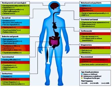
Figure 1: Williams’ Syndrome - myriads of clinical features

pedia_751_02.JPG
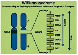
Figure 2: There is microdeletion on chromosome 7, resulting in the loss of 26-28 genes.

The neurocognitive profile of William's syndrome most commonly includes mild mental retardation. Cognitive strengths and weaknesses are relative to other patients with mental retardation consist of relatively good auditory rote memory but extreme difficulty with visuospatial construction tasks [2]. Most of the patients with WS have CHDs, which typically include supravalvular aortic stenosis and/or supravalvular pulmonary stenosis [3, 4]. Patients with WS may commonly develop hypertension either because of renal artery stenosis

or COA or other undefined etiologies [3]. Approximately 90% of WS have a deletion at chromosome 7q11.23, which can be detected by FISH (fluorescent in situ hybridization) (Figure 2).

The genes mapping to this region have been defined and include the elastin gene. In isolated supravalvular aortic stenosis [5, 6] association of deletion of the elastin gene in patients with WS is thought to account for the cardiovascular phenotype [7].

Case Report

A two-month-old female infant was referred to us for comprehensive echocardiographic evaluation along with clinical evaluation and management of severely dyspnoeic children. The parents were especially cooperative while enumerating the details of the history. The child was a full-term normal delivery from a multipara woman of 32 years and they denied any family history of congenital heart disease. The mother and child were appropriately vaccinated at the time of presentation. Moreover, there was no history of maternal risk factors for CHD (morbid obesity, diabetes, febrile illness, smoking, alcohol intake, teratogenic drug usage or radiation exposure). According to the parents, the child was normal till 2 weeks in the postnatal period. Then she started developing a cough, breathlessness, chest rétractions and failure to thrive.

pedia_751_03.JPGFigure 3: severely dyspneic, irritable child, with obvious chest retractions and pectus excavatum deformity.


On clinical examination, the child was of average build, severely dyspnoeic, highly irritable, with rapid intercostal retractions and mild sweating over the forehead (Figure 3).

There was no evidence of clubbing or cyanosis. The child was weighting 2.2 kg, height of 35cm, had a respiratory rate of 35/mm, and SPO2 was 91% at room air. The blood pressure was 130/80 mmHg in the right upper extremity and 98/60 in the right lower extremity, indicating the presence of systemic hypertension and moderate coarctation of the aorta.

Distinctive "Elfin-like” facies was evident (Figure 4), with the presence of:

pedia_751_04.JPGFigure 4: Typical Elfin facies: wide forehand, bilateral epicanthal folds, wide-set eyes, sunken bridge of the nose, wide nostrils with upturned nose, full cheeks, pointed chin, large right ear, left ear having cup deformity.

i. Prominent wide forehand

ii. Full cheeks

iii. Sunken nasal bridge

iv. Long upper lip length (philtrum)

v. Upturned nose

vi. Small chin

vii. Wide-set eyes

viii. The large right external ear

ix. Bilateral epicanthal folds

x. Cup ear deformity of the left ear

xi. Puffiness around the eyes

xii. Full cheeks

Besides the conspicuous facies and the presence of pectus excavatum, no other musculoskeletal anomalies were identified. There was the presence of bilateral scattered crept in the interscapular, base and axillary region of the chest.

On cardiovascular examination, a grade 3/6 ejection systolic murmur was audible over the precordium, best heard in the left second intercostal space, just adjacent to the sternum and also in the left infraclavicular region. No radiation was detected in the carotids. All the peripheral arteries were normally palpable.

pedia_751_05.JPG
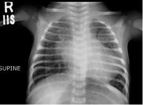
Figure 5: Chest Xray PA view Cardiomegaly, Biatrial enlargement, Pulmonary Venous congestion, Pulmonary

pedia_751_06.JPG

Figure 6: Resting 12 lead ECG shows sinus tachycardia, with a rate of 136/ min. No other abnormalities were appreciated.

X-ray chest PA view (Figure 5) showed gross cardiomegaly, pulmonary venous congestion and pulmonary edema. Resting 12 lead ECG revealed (Figure 6), sinus tachycardia with a heart rate of 136/min with no evidence of any chamber enlargement, arrhythmia or A-V blocks.


Pathological tests were unremarkable except for elevated levels of total serum calcium and serum ionic calcium (13.9 mg/dl and 7 mg/dl respectively), suggestive of hypercalcemia. We want to highlight that no genetic studies were done, because of the non-availability of the FISH technique.

Comprehensive Color Doppler Echocardiography

The patient underwent colour echocardiography and a detailed sequential chamber analysis was performed by the author with the patient lying in a supine and left lateral decubitus position. Imaging was done from the subcostal, parasternal long axis (LX), parasternal short axis (SX) apical 4-chamber (4CH), apical 5-chamber (5CH) and suprasternal views.

There was levocardia, situs solitus, atrioventricular concordance, ventricular-arterial concordance, concordant d- bulbo-ventricular loop, normally related great arteries & and left aortic arch. Systemic and pulmonary venous drainage was normal. In the SX view (Figure 7), a constriction band is visualized, just above the bifurcation of the main pulmonary. The peak gradient and mean gradient across the stenotic band was 18.3/10.3 mmHg (Figure 8), signifying a mild gradient. Nonetheless, the pulmonary valve (PV) annulus and main pulmonary artery (MPA) were dilated (annulus (D) 14.2 mm MPA (D) 13.4mm).

pedia_751_07.JPG
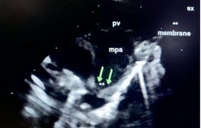
Figure 7: supravalvular pulmonary stenosis- In the SX view, a constriction band is visualised just above the bifurcation of the main pulmonary artery. The pulmonary valve annulus and main pulmonary artery are dilated and the left and right branch pulmonary arteries are of normal size: pv annulus (D) 14.2 mm, main pulmonary artery (D) 13.4 mm, left pulmonary artery (D) 4.9 mm, right pulmonary artery (D) 4.6 mm

pedia_751_08.JPG
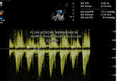
Figure 8: supravalvular pulmonary stenosis: On CW analysis peak/mean gradient across the supravalvular stenotic band was 17.7/9 mmhg, corresponding with mild supravalvular stenosis

The left pulmonary artery (LPA) and right pulmonary artery (RPA) were normal (LPA (D) 4.9 mm, RPA (D) 4.6mm). On CW Doppler analysis peak/mean gradient across the supravalvular stenotic band was 17.7/9 mmhg, corresponding with mild supravalvular stenosis( Figure 8).

Additionally, a moderate-sized PDA (size: 5mm) was identified, as a turbulent red-coloured jet in the pulmonary artery (Figure 9) with the left to right shunt. The peak and mean gradient across PDA was 40.5/14.5 mmHg (Figure 10).

pedia_751_09.JPG
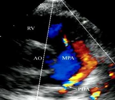
Figure 9: PDA - In the SX view on color flow doppler analysis a continuous flow (red) from the PDA into the main pulmonary artery is displayed. AO, aorta, RV, right ventricle, MPA, main pulmonary artery


pedia_751_10.JPG
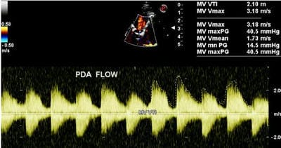
Figure 10: On CW Doppler analysis across PDA peak/mean gradient was 40.5/ 14.5 MMHg

In the suprasternal view, a distinct COA is visualized, just distal to the origin of the left subclavian artery (Figure 11).

pedia_751_11.JPG
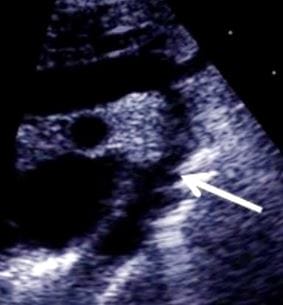
Figure 11: suprasternal view: The arrow depicts the site of COA, just beyond the origin of the left subclavian artery.

On colour echocardiography a turbulent mosaic pattern flow across the COA is demonstrated (Figure 12).

On CW Doppler analysis a peak/mean gradient across the COA was 43.5/25 mmHg (Figure 13) consistent with moderate COA.

There was no evidence of aortic hypoplasia at the ascending, arch and descending thoracic aorta level.

pedia_751_12.JPG
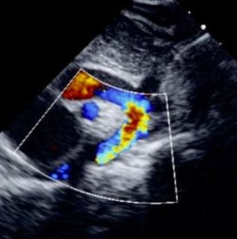
Figure 12: Suprasternal View demonstrates a turbulent mosaic pattern flow in the region of COA and descending aorta.

pedia_751_13.JPG
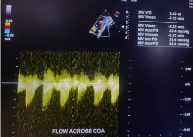
Figure 13: CW Doppler flow across COA is indicative of a peak/ mean gradient of 43.5/25 mm hg, consistent with moderate COA

In the 4CH view, there was the presence of tricuspid regurgitation (TR) with a central jet occupying 25% of the right atrial area (Figure 14).

On CW Doppler analysis of the TR jet (Figure 15), the peak velocity was 3.6 m/sec (peak gradient 52 mmHg).

The estimated right ventricular systolic pressure (RVSP) pulmonary artery pressure (PAP) was 67 mmHg, suggestive of severe pulmonary hypertension.


pedia_751_14.JPG
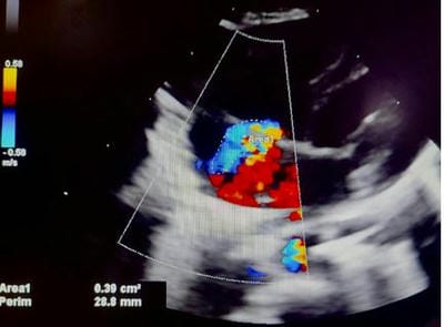
Figure 14: 4CH View: A turbulent, moderate tricuspid regurgitation jet (blue) is recognized in the right atrium.

pedia_751_15.JPG
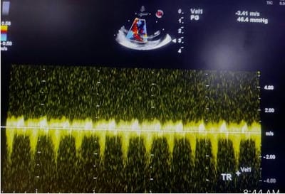
Figure 15: CW Doppler across Tricuspid Regurgitation jet shows a peak velocity of 3.6 m/ sec (gradient 52 mm hg). The estimated right ventricular systolic pressure/ pulmonary artery pressure was (52+ 15) 67 mm hg, suggestive of severe pulmonary hypertension.

pedia_751_16.JPG
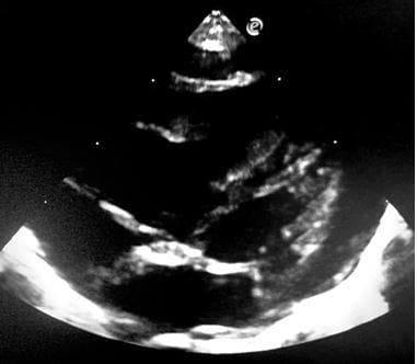
Figure 16: modified 4CH view: delineates dilatation of right ventricular and right atrium.

Correspondingly, there was dilatation of the right ventricle (RV) and right atrium (RA) (Figure 16). Additionally, there was mild dilatation of the left atrium and left ventricle with a normal biventricular systolic function. LVEF was 77%.

Considering the current life-threatening situation of the infant, because of the presence of ongoing heart failure and the complexity of the congenital heart defects, we suggested IV prostaglandins to keep the PDA open, along with guideline-based anti-congestive medical therapy. Furthermore, we emphasized the need for urgent surgical intervention for the correction of COA, PDA and SVPS and hence the patient was referred to a tertiary care pediatric cardiovascular institute.

Discussion

WS is a complex developmental disorder characterized by congenital cardiovascular disorder mental retardation characteristic learning profile, a hypersocial personality, and infantile hypercalcemia [1].

Although cardiovascular disease is one of the most common presentations of WS, it is rarely associated with significant morbidity and mortality in the neonatal and infantile periods.

Its prevalence has been estimated to range from 1:13,700- 1:25,000 live births [1]. This syndrome is associated with a microdeletion in the chromosome region 7q11.23, encompassing the elastin gene. Reports suggest that this microdeletion of the elastin gene is responsible for typical vasculopathy of WS, supravalvular aortic stenosis (SVAS) and pulmonary artery stenosis [2, 3].

Cardiovascular abnormalities occur in approximately 80% of reported cases with SVAS being a common cardiac anomaly, present in 64% of patients. Other lesions include pulmonary artery stenosis (13%), aortic hypoplasia, coarctation of the aorta (COA 8%), mitral valve prolapse (15%), and sepal defects [1]. SVAS usually progresses with age, whereas pulmonary artery stenosis decreases with increasing age.

Moreover, intellectual disability with a peculiar cognitive and/or behavior profile, characteristic facies and occasional hypercalcemia are also described [8]. The definite diagnosis should be made by the clinical picture assessed by a medical


geneticist together with a demonstration of the typical elastin gene hemizygosity (assessed by FISH); however, genetic testing is often unavailable in developing countries including India. Therefore the combination of a typical clinical phenotype and echocardiographic profile could help to confirm the diagnosis.

Our patient had hypercalcemia and multiple facial features consistent with “Elfin-like” facies as documented by numerous earlier studies.

The diagnosis of SVAS and other vasculopathy like COA and pulmonary artery stenosis can be made by multiple imaging modalities. The defining feature of this malformation is an aortic narrowing at the level of the simotubuar junction, but in some cases, there is a narrowing of the entire arch and abdominal aorta. In our patient, there was the presence of moderate COA and mild supravalvular pulmonary stenosis as shown by other authors [9]. SVAS often presents in childhood and if not corrected by surgery can lead to heart failure and death [10], Although SVAS was not present in our index patient the existence of a constellation of moderate COA, systemic hypertension of 130/80 mmHg and moderate-sized PDA could have resulted in rapid progression of heart failure. This persistently very high overload could have produced early and refractory symptoms. There are case reports of WS with rapid progression of COA and severe middle aortic syndrome which required corrective surgery within 2 months of life [11, 12].

Conclusion

Cardiovascular abnormalities are present in the large majority of patients with Williams Syndrome, and the need for medical or surgical interventions is common. Although cardiovascular presentation of WS, is rarely associated with significant morbidity and mortality in the neonatal and infantile period. Advancements in surgical techniques may provide options for significant improvements in our patient with WS presenting with multiple CHDS, severe pulmonary hypertension and heart failure.

Reference

Moris CB, Mervis CA. Wiliams syndrome and related disorders. Annual Review of Genomics and Human Genetics. 2000; 1:461-484. [Crossref][PubMed][Google Scholar]

Mervis CB, Robinson BF, Bertrand J, Morris CA, Klein-Tasman BP, Armstrong SC. The Williams syndrome cognitive profile. Brain Cogn. 2000;44(3):604-628. doi:10.1006/brcg.2000.1232 [Crossref][PubMed][Google Scholar]

Kececioglu D, Kotthoff S, Vogt J. Williams-Beuren syndrome: a 30-year follow-up of natural and postoperative course. Eur Heart J. 1993;14(11):1458-1464. doi:10.1093/eurheartj/14.11.1458 [Crossref][PubMed][Google Scholar]

Eronen M, Peippo M, Hiippala A, et al. Cardiovascular manifestations in 75 patients with Williams syndrome. J Med Genet. 2002;39(8):554-558. doi:10.1136/jmg.39.8.554 [Crossref][PubMed][Google Scholar]

Curran ME, Atkinson DL, Ewart AK, Morris CA, Leppert MF, Keating MT. The elastin gene is disrupted by a translocation associated with supravalvular aortic stenosis. Cell. 1993;73(1):159-168. doi:10.1016/0092-8674(93)90168-p [Crossref][PubMed][Google Scholar]

Metcalfe K, Rucka AK, Smoot L, et al. Elastin: mutational spectrum in supravalvular aortic stenosis. Eur J Hum Genet. 2000;8(12):955-963. doi:10.1038/sj.ejhg.5200564 [Crossref][PubMed][Google Scholar]

Frangiskakis JM, Ewart AK, Morris CA, et al. LIM-kinase1 hemizygosity implicated in impaired visuospatial constructive cognition. Cell. 1996;86(1):59-69. doi:10.1016/s0092-8674(00)80077-x [Crossref][PubMed][Google Scholar]

Morris CA, Demsey SA, Leonard CO, Dilts C, Blackburn BL. Natural history of Williams syndrome: physical characteristics. J Pediatr. 1988;113(2):318-326. doi:10.1016/s0022-3476(88)80272-5 [Crossref][PubMed][Google Scholar]

Sugayama SM, Moisés RL, Wagënfur J, et al. Williams-Beuren syndrome: cardiovascular abnormalities in 20 patients diagnosed with fluorescence in situ hybridization. Arq Bras Cardiol. 2003;81(5):462-473. doi:10.1590/s0066-782x2003001300003 [Crossref][PubMed][Google Scholar]

Keating MT. Genetic approaches to cardiovascular disease. Supravalvular aortic stenosis,


Williams syndrome, and long-QT syndrome. Circulation. 1995;92(1):142-147. doi:10.1161/01.cir.92.1.142 [Crossref][PubMed][Google Scholar]

Monfared A, Messner A. Death following tonsillectomy in a child with Williams syndrome. Int J Pediatr Otorhinolaryngol. 2006;70(6):1133-1135. doi:10.1016/j.ijporl.2005.11.009 [Crossref][PubMed][Google Scholar]

Hall EK, Glatz J, Kaplan P, et al. A case report of rapid progressive coarctation and severe middle aortic syndrome in an infant with Williams syndrome. Congenit Heart Dis. 2009;4(5):373-377. doi:10.1111/j.1747-0803.2009.00287.x [Crossref][PubMed][Google Scholar]