E-ISSN:2349-3267
P-ISSN:2349-5499

Review Article

Maternal Serum

Pediatric Review - International Journal of Pediatric Research

2024 Volume 11 Number 2 Apr-Jun
Publisherwww.medresearch.in

Association of Neural Tube Defects of Newborn with Maternal Serum Vitamin-B12 level-A Review Study

Badal F1*, Rashid F2, Pervin R3, Naher N4, Khanam M5
DOI:https://doi.org/10.17511/ijpr.2024.i02.01

1* FA Badal, Assistant Professor, Department of Neonatology, Shaheed Ziaur Rahman Medical College, Bogura, Bogra, Bangladesh.

2 FJB Rashid, Assistant Surgeon, Obstetrics and Gynecology, Sheikh Hasina Medical College Hospital, Tangail, Bangladesh.

3 R Pervin, Assistant Professor, Obstetrics and Gynecology, Sheikh Hasina Medical College, Tangail, Bangladesh.

4 N Naher, Junior Consultant, Gynae and Obs, Bedded General Hospital, Tangail, Bangladesh.

5 M Khanam, Junior Consultant, Gynae and Obs, Sheikh Hasina Medical College Hospital, Tangail, Bangladesh.

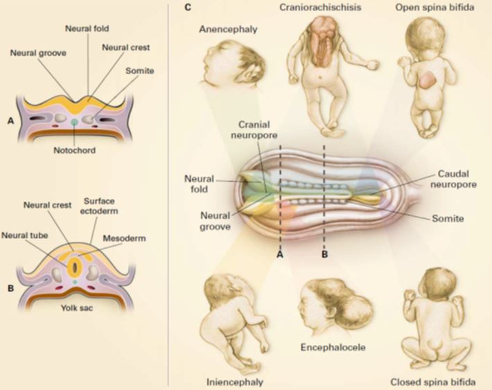
Neural tube defects (NTDs) are one of the most common birth defects in our clinical practices. Maternal nutritional factors are associated with an increased chance of development of NTDs in offspring. Periconceptional folic acid supplementation can prevent up to three-fourths of neural tube defects (NTDs). Some studies reported that there might be an association between maternal serum vitamin B12 levels and NTDs. Dietary vitamin B12 deficiency is a common problem in the Indian subcontinent including Bangladesh because of inadequate dietary intake, dietary habits and/or malabsorption. There is a scarcity of studies to establish the relationship between vitamin B12 and NTDs. It would be beneficial to see the association of vitamin B12 and NTDs in a Bangladeshi population, which may help in deciding about vitamin B12 supplementation as a preventive measure of NTDs.

Keywords: NTDs, Maternal and Neonatal Serum, vitamin B12 level

Corresponding Author How to Cite this Article To Browse
FA Badal, Assistant Professor, Department of Neonatology, Shaheed Ziaur Rahman Medical College, Bogura, Bogra, Bangladesh.
Email:
Badal F, Rashid F, Pervin R, Naher N, Khanam M, Association of Neural Tube Defects of Newborn with Maternal Serum Vitamin-B12 level-A Review Study. Pediatric Rev Int J Pediatr Res. 2024;11(2):7-14.
Available From
https://pediatrics.medresearch.in/index.php/ijpr/article/view/765

Manuscript Received Review Round 1 Review Round 2 Review Round 3 Accepted
2024-05-16 2024-05-22 2024-05-28 2024-06-03 2024-06-09
Conflict of Interest Funding Ethical Approval Plagiarism X-checker Note
Nil None yes 13.32

© 2024by Badal F, Rashid F, Pervin R, Naher N, Khanam Mand Published by Siddharth Health Research and Social Welfare Society. This is an Open Access article licensed under a Creative Commons Attribution 4.0 International License https://creativecommons.org/licenses/by/4.0/ unported [CC BY 4.0].

Introduction

Neural tube defects (NTDs) are a group of serious birth defects that arise when the neural tube fails to develop into the brain and spinal cord during the first month of pregnancy. The neural tube usually closes by day 28 of prenatal life, and its incomplete or incorrect closure results in malformations called neural tube defects (NTDs) [1]. There is a spectrum of severity of NTDs from anencephaly, which is incompatible with life beyond the neonatal period, to spina bifida occulta, which may be asymptomatic (. In the United States, the incidence of NTDs was estimated at 1 per 1,000 deliveries, anencephaly at 0.6-0.8 per 1,000 live births, and open spina bifida at 0.5-0.8 per 1,000 live births [2].

The incidence varies from 1/100 live births in certain regions of China to about 1/5000 live births in Scandinavian countries. In Bangladesh prevalence of NTDs is 4.7 per thousand live births. Some hospital-based studies showed NTDs were found in 13.33% and 11.32% [3] of total birth defects enrolled on their studies. The etiology of NTDs is considered multifactorial, with genetic, environmental and nutritional factors playing some role [4].

The nutritional status of pregnant women may influence the vulnerability to NTDs in the fetus. The search for other etiological factors, particularly nutritional factors, has been continuing. Maternal nutritional risk factors are associated with an increased likelihood of the development of NTDs in offspring including folate deficiency in the periconceptional period, and low maternal vitamin B12. Folic acid can prevent up to three-fourths of neural tube defects (NTDs). Some studies suggest that nutrients other than folate may also be essential to neural tube closure and have a potential role in risk reduction, with vitamin B12 mentioned most often [5].

Some studies showed low maternal vitamin B12 levels increase the risk of NTD. Vitamin B12 is a necessary cofactor for two enzymes in DNA synthesis: folate-dependent methionine synthesis and folate–independent methyl malonyl CoA mutase. Maternal serum vitamin B12 status before and during pregnancy is strongly associated with cord blood concentrations of vitamin B-12 and total homocysteine as well as infant stores of the vitamin at birth.

Moreover, fetal vitamin B12 accumulation during gestation is the major determinant of the B12 status of the newborn and is stored in infancy. Therefore, this hospital-based study was conducted to see the association of NTDs with maternal and neonatal serum vitamin B12 levels.

NTDs with maternal and neonatal serum vitamin B12 level

NTDs represent one of the most common congenital malformations in neonates worldwide. They constitute a heterogeneous group of disorders that occur during the first few weeks of gestation involving specific elements of the neural tube and its derivatives [6].

The central nervous system (CNS) appears at the beginning of the third week as a slipper-shaped plate of thickened ectoderm, the neural plate, in the mid-dorsal region in front of the primitive node. Its lateral edges soon elevate to form the neural folds.

Pathophysiology

Two distinct processes appear to be involved in the formation of the neural tube: primary neurulation and secondary neurulation (i.e.canalization). The neural plate and the notochord are formed during early embryonic development. The neural groove develops by the third gestational week. Subsequently, the neural folds form bilaterally.

pediatric_765_01.JPG
Figure 1: Pathophysiology

Features of Neural-Tube Development and Neural-Tube Defects [7]. Panel A shows a cross-section of the rostral end of the embryo at approximately three weeks after conception, showing the neural groove in the process of closing, overlying the notochord.


The neural folds are the rising margins of the neural tube, topped by the neural crest, and demarcate the neural groove centrally. Panel B shows a cross-section of the middle portion of the embryo after the neural tube has closed. The neural tube, which will ultimately develop into the spinal cord, is now covered by surface ectoderm (later, the skin). The intervening mesoderm will form the bony spine.

The notochord is regressing. Panel C shows the developmental and clinical features of the main types of neural tube defects. The diagram in the centre is a dorsal view of a developing embryo, showing a neural tube that is closed in the centre but still open at the cranial and caudal ends.

The dotted lines marked A and B refer to the cross sections shown in Panels A and B. Shaded bars point to the region of the neural tube relevant to each defect. In anencephaly, the absence of the brain and calvaria can be total or partial.

Genetic disorders

While some neural tube defects may arise as a result of gene-environment interaction, several identified syndromes also can give rise to neural tube defects. Some 60 syndromes that have a neural tube defect component together with other birth defects have been identified.

Each of the different types of neural tube defects may arise as a result of one or more of these syndromes. There is also evidence that non-syndrome neural tube defects may sometimes also have a genetic source, based on familial and twin studies.

Environmental exposures

Since neural tube defects are rare birth defects, it is hard to definitively associate an environmental exposure to a specific N'I'D. However, several studies have identified exposures that may cause neural tube defects. A selection of these is listed in Table 1, to give an idea of the range of exposures in the first trimester of pregnancy that may give rise to neural tube defects.

Occupational exposure to glycol ethers, pesticides, drinking water (hard water), cured meat, and blighted potatoes containing high concentrations of nitrates and lead are among the important exposures found to be associated with NTD. Maternal tea consumption has also been described in the etiology of NTD [8].

Maternal factors

General conditions

Several maternal conditions may contribute to an increased risk of an NTD-affected pregnancy. It was found that the incidence of NTD among infants of women who had a fever in the 1SI trimester was higher than control afebrile women. However, it was not clear whether the exposure to high temperatures or the bacteria or virus was the cause of the maldevelopment of the neural tube [9].

Women with diabetes were found to be four times associated with pregnancies involving neural tube defects (95 percent confidence interval [3.1, 5.1]) in comparison to non-diabetics. However, the investigators found a much smaller odds ratio of 1.3 (95 percent confidence interval [1.0, 1.6]) of neural tube defect pregnancies among women with gestational diabetes.

Obese women were found to be 3.5 times more, affected by NTD in comparison to non-obese. Maternal psychological stress may increase the risk of neural tube defects. It was reported that women who experienced the death of someone close, job loss, separation or divorce - either themselves or someone close to them in the very early stages of pregnancy had an odds ratio of 1.5 (95 percent confidence interval [1.1, 2.1]) [10].

Maternal age

The literature reports associations between maternal age and the incidence of neural tube defects. Prevalence of NTD-affected pregnancies was found to be decreased with increasing maternal age. Meanwhile, some other authors have found a U-shaped distribution of neural tube defects in relation to maternal age.

Maternal diet

Folic acid is a nutrient that is needed for proper neural tube development. In 1992, the U.S. Centers for Disease Control and Prevention recommended that all women of childbearing age consume 0.4 mg of folic acid daily.

This routine supplementation prevents the occurrence of NTD by more than 50% when it is taken before conception and continued throughout the first trimester of pregnancy. For women who have had a child with an NTD the recommendation for daily consumption of folic acid is 4000 µg.


This supplementation in addition to folate in the diet before and during early pregnancy resulted in a 71% reduction of recurrence of NTDs. The recommendation originated in research indicating that -inadequate levels of folic acid in the first weeks of pregnancy increased the risk of having a baby with a neural tube defect. In 1996, the U.S. Food and Drug Administration agreed that enriched cereal-grain products must be fortified with folic acid, as a means to reduce the rate of neural tube defects in the United States. There is evidence to suggest that excess lead (Pb) is causally related to NTD especially anencephaly [11,12].

Besides, deficiencies of vitamin B12, zinc, selenium and have been described to be associated with increased incidence of NTD [13].

Vitamin B12 and Neural Tube Defects

Vitamin B12 deficiency was first described in 1849 and was considered to have a fatal outcome until 1926 when a diet of liver, high in vitamin B12, was shown to slow the disease process.

Function

Vitamin B12 also known as cobalamin, comprises several forms including cyano-, methyl-,deoxyadenosyl- and hydroxy-cobalamin. The cyano form, which is used in supplements, is found in trace amounts in food [14]. The other forms of cobalamin can be converted to the methyl- or 5-deoxyadenosyl forms that are required as co-factors for methionine synthase and L-methyl-malonyl-CoA mutase.

Methionine synthase is essential for the synthesis of purines and pyrimidines. The reaction depends on methylcobalamin as a co-factor and is also dependent on folate, in which the methyl group of methyltetrahydrofolate is transferred to homocysteine to form methionine and tetrahydrofolate. Therefore vitamin B12 is also necessary for erythropoiesis and essential for normal neurodevelopment [15].

Food Sources

Vitamin B12 is synthesized by certain bacteria in the gastrointestinal tract of animals and is then absorbed by the host animal.

Vitamin B12 is concentrated in animal tissues, hence, vitamin B12 is found only in foods of animal origin [16].

Foods that are high in vitamin B12 include liver, kidney, beef and lamb, chicken, eggs, fish and milk, curd and dairy foods.

Recommended dietary allowance (RDA)

3 µg for adults, o.5-1.5 µg for children and 4 µg for pregnancy and lactation.

Absorption

Vitamin B12 from dietary intake is bound to proteins and is released by hydrochloric acid that is secreted by the parietal cells from the stomach. The free form of vitamin B12 is then immediately bound to haptocorrins, which are glycoproteins secreted by the stomach and salivary glands. These haptocorrins protect vitamin B12 from chemical denaturation in the stomach. The parietal cells secrete an intrinsic factor that binds to vitamin B12 and enables its active absorption.

When the contents of the stomach enter the duodenum, the haptocorrins become partly digested by the pancreatic proteases. This causes them to release their vitamin B12. Immediately after they are released, they get bound to intrinsic factors in the duodenum. The vitamin B12-intrinsic factor complex then proceeds to the ileum, where it is absorbed by specific ileal receptors [17].

Metabolic reaction

pediatric_765_02.JPG
Figure 2:
The Metabolic Roles of Folic Acid and vitamin B12

After entering the cell, folic acid, possibly with the aid of folate receptors, is involved in the transfer of carbon atoms that are used to synthesize nucleotides or, through the conversion of homocysteine to methionine, for the methylation of a variety of substrates. These processes are regulated by numerous molecules, including enzymes and vitamins other than folic acid (e.g., vitamins B6 and B12).


The activity of some enzymes, such as methionine synthase, may be influenced by other enzymes, such as methionine synthase reductase. Enzymes and factors that may be important in neural tube development are indicated by boxes. Cobalamin is involved as a coenzyme in 2 enzymatic reactions.

In the first enzymatic reaction, methionine synthesis produces methionine from homocysteine in the cytosol. In the second enzymatic reaction, adenosylcobalamin is involved as a coenzyme in the production of methylmalonyl-co-enzym-A (methylmalonyl-CoA) to succinyl-CoA in the mitochondrion.

A vitamin B12 deficiency results in elevated concentrations of homocysteine and methylmalonic acid in serum [17].

Vitamin B12 deficiency and pregnancy

The reported prevalence of deficient plus low serum B-12 concentrations reported in Latin America is typically around 40% and reaches 60 to 80% in some populations in Africa and Asia, affects all ages, and is associated with low animal source food consumption [18].

From the first through the third trimester of pregnancy, there is a gradual decline in the serum concentration of vitamin B12 due to hemodilution, hormonal changes, alterations in the concentration of vitamin B12 binding proteins, and placental transport of vitamin B12 to the fetus.

The lowest concentration of vitamin B12 is seen at 32 weeks of pregnancy and before delivery, it increases again to reach a normal level after birth. The concentration of the active part of vitamin B12, holotranscobalamin, remains unchanged during pregnancy [19].

Types of Neural Tube Defects

The main types of neural tube defects (NTDs) are described below. The corresponding codes from the International Classification of Diseases -Ninth Revision Clinical Modification (ICD-9-CM) also are provided.

Anencephaly

Anencephaly (1CD-9-CM 740.0) occurs when the cephalic end of the neural tube fails to close, resulting in the absence of a major portion of the brain, skull and scalp.

Many infants with anencephaly are delivered stillborn; those born alive are usually blind, deaf, unconscious and unable to feel pain. If the infant is not stillborn, then he or she will usually die within a few hours or days after birth.

Antenatal diagnosis and preventive aspects

The incidence of NTDs is declining throughout the world following the introduction of ultrasound examination and measurement of maternal serum alpha-fetoprotein (MSAFP), amniotic fluid alpha-fetoprotein (AFAFP) and amniotic fluid acetylcholinesterase (AFAChE) [20]. MSAFP is widely offered as a screening test for the detection of NTDs in pregnancy in addition to a careful examination of the fetal brain and spine with ultrasound during an anomaly scan at 18-20 weeks gestation. USG is used both as a screening test and as a follow-up test after positive results of an MSAFP screening. AFAFP and AFAChE are employed primarily as a confirmation test [20].

Clinical assessment

Examination of the newborn baby should be performed by a clinician with appropriate experience. The width and length of the spinal lesion should be measured and its position relative to the spinal column recorded. Within the cystic lesion is the neural plaque, the maldeveloped spinal cord that is exposed on the surface.

Immediately after birth, the nerve roots can be seen through the CSF within the sac: they usually run horizontally or even upward because the physiological cranial migration of the cord was impeded during fetal life. The neural plaque may occupy the centre or the upper or lower part of the whole lesion. A large plaque with a high upper level carries a bad prognosis, with extensive paralysis and analgesia [2].

Paralysis

There is a wide range of paralysis possible in the legs, from none to complete. Typically, the paralysis is of lower motor neuron type (flaccid) [2]. Knowledge (and a chart) of the segmental innervations of the leg muscles is helpful and the neurological level of paralysis may be ascertained in each leg separately and charted. The neurological level does not necessarily coincide with the vertebral level. All muscles below the uppermost affected segment may be paralyzed [21].


Another pattern also occurs, especially in thoracolumbar lesions, with flaccid paralysis of muscles deriving their innervations from the affected segments followed by reflex movements in muscles below the affected segments [22].

Reflex activity, which later disappears and may be followed by spasticity, should not be mistaken for useful voluntary movement. Stimulation of the legs may provoke reflex movements and mislead the examiner; it is better to stimulate the upper half of the body to elicit active movement in unaffected muscle groups.

The sphincters and the urinary tract

Almost all babies with myelomeningocele have paralysed urethral sphincters [2].

In the absence of normal sphincter control, urine can be expressed. An intravenous urethrogram a renal ultrasound and a micturating cystourethrogram will show whether there is hydronephrosis, retention of urine, trabeculation or diverticula in the bladder and/or ureteric reflux.

The cxprcssibility of the bladder and the residual urine can also be demonstrated. Other renal defects, such as single kidney, horseshoe kidneys; pelvic kidney and double pelvis and ureters are some ten times more commoner than in infants without NTDs.

The anus is not visible in normal babies without separating the buttocks, but when the muscles of the perineum and the rectal sphincters are . paralysed it is on the surface and patulous.

Perianal rugae may still be present. The anal sphincter cannot contract on perianal stimulation or grip a small finger inserted into the rectum.

Hydrocephalus

The maximum head circumference must be measured and charted and related to the baby's weight and gestational age.

Cranial ultrasound is an essential early investigation in myelomeningocele and can reveal dilated ventricles before the head circumference enlarges.

Ultrasound, computed tomography (CT) or MR imaging can also reveal the associated Chiari malformation, which can cause bulbar paresis. Treatment is the insertion of a ventricular shunt.

Diagnostic evaluation

During the first hospitalization, the following tests should be done on most children with meningomyelocele. Scheduling these tests will vary depending on each situation [23].

Biochemical analysis

A 2- 3 ml blood sample was obtained from each subject of both groups by venipuncture and collected in capped polypropylene tubes labelled with the allocated number. The serum was separated by centrifugation (4000 rpm for 5 minutes), and the haemolysed sample was discarded. The level of serum vitamin B12 was expressed in pgm/ml. Cut-off values for B12 represented deficiency (<200 pg/ml), marginal deficiency (200-300 pg/ml), and normal status (>300 pg/ml) [24].

Conclusion

Maternal nutritional deficiencies can increase chance of neural tube defects (NTDs)of newborn. Folate deficiency can increase the probability of a fetus to have NTDs as compared to pregnancies with normal folate.Besides this maternal vitamin B12 deficiency have risk of NTDs of a newborn than normal vitamin B12 level. This review study focuses attention to the importance of nutritional factors in the etiology of neural tube defects.

References

1. Green NS. Folic acid supplementation and prevention of birth defects. J Nutr. 2002 Aug;132(8 Suppl):2356S-2360S. doi: 10.1093/jn/132.8.2356S. PMID: 12163692 [Crossref][PubMed][Google Scholar]

2. Rennie, JM (ed. ) 2012, Rennie and Roberton’s Textbook of Neonatology, 5th edn, Churchill Livingstone Elsevier, London. . . doi: 10.1093/jn/132.8.2356S. PMID: 12163692 [Crossref][PubMed][Google Scholar] [Crossref][PubMed][Google Scholar]

3. Islam, MN, Siddika, M, Bhuiyan, MKJ, & Chowdhury, AM 2013, ‘Pattern of Congenital Anomalies in Newborns in a Tertiary Level Hospital in Bangladesh’, Journal of Surgery Pakistan (International), vol. 18, no. 1, pp. 32-35. [Crossref][PubMed][Google Scholar]


4. Harris MJ, Juriloff DM. Mini-review: toward understanding mechanisms of genetic neural tube defects in mice. Teratology. 1999 Nov;60(5):292-305. doi: 10.1002/(SICI)1096-9926(199911)60:5<292::AID-TERA10>3.0.CO;2-6. PMID: 10525207 [Crossref][PubMed][Google Scholar]

5. Schorah CJ, Smithells RW, Scott J. Vitamin B12 and anencephaly. Lancet. 1980 Apr 19;1(8173):880. doi: 10.1016/s0140-6736(80)91381-1. PMID: 6103238 [Crossref][PubMed][Google Scholar]

6. Cloherty, JP, Eichenwald, EC, Hansen, AR, & Stark, AR (eds. ) 2012, Manual of neonatal care, 7th edn ,Wolter Kluwer & Lippincott Williams and Wilkins, Philadelphia. . . doi: 10.1016/s0140-6736(80)91381-1. PMID: 6103238 [Crossref][PubMed][Google Scholar] [Crossref][PubMed][Google Scholar]

7. Botto LD, Moore CA, Khoury MJ, Erickson JD. Neural-tube defects. N Engl J Med. 1999 Nov 11;341(20):1509-19. doi: 10.1056/NEJM199911113412006. PMID: 10559453 [Crossref][PubMed][Google Scholar]

8. Correa A, Stolley A, Liu Y. Prenatal tea consumption and risks of anencephaly and spina bifida. Ann Epidemiol. 2000 Oct 1;10(7):476-477. doi: 10.1016/s1047-2797(00)00144-7. PMID: 11018418 [Crossref][PubMed][Google Scholar]

9. Shaw GM, Todoroff K, Velie EM, Lammer EJ. Maternal illness, including fever and medication use as risk factors for neural tube defects. Teratology. 1998 Jan;57(1):1-7. doi: 10.1002/(SICI)1096-9926(199801)57:1<1::AID-TERA1>3.0.CO;2-6. PMID: 9516745 [Crossref][PubMed][Google Scholar]

10. Grewal J, Carmichael SL, Song J, Shaw GM. Neural tube defects: an analysis of neighbourhood- and individual-level socio-economic characteristics. Paediatr Perinat Epidemiol. 2009 Mar;23(2):116-24. doi: 10.1111/j.1365-3016.2008.00992.x. PMID: 19159398; PMCID: PMC2865191 [Crossref][PubMed][Google Scholar]

11. Bound JP, Harvey PW, Francis BJ, Awwad F, Gatrell AC. Involvement of deprivation and environmental lead in neural tube defects: a matched case-control study. Arch Dis Child. 1997 Feb;76(2):107-12. doi: 10.1136/adc.76.2.107. PMID: 9068297; PMCID: PMC1717078 [Crossref][PubMed][Google Scholar]

12. McMichael AJ, Dreosti IE, Ryan P, Robertson EF. Neural tube defects and maternal serum zinc and copper concentrations in mid-pregnancy: a case-control study. Med J Aust. 1994 Oct 17;161(8):478-82. doi: 10.5694/j.1326-5377.1994.tb127560.x. PMID: 7935120 [Crossref][PubMed][Google Scholar]

13. McMichael AJ, Dreosti IE, Ryan P, Robertson EF. Neural tube defects and maternal serum zinc and copper concentrations in mid-pregnancy: a case-control study. Med J Aust. 1994 Oct 17;161(8):478-82. doi: 10.5694/j.1326-5377.1994.tb127560.x. PMID: 7935120 [Crossref][PubMed][Google Scholar]

14. Scott, JM 1997,’Bioavailability of vitamin B12’, European Journal of Clinical Nutrition, vol. 51, pp. 49-53. . [Crossref][PubMed][Google Scholar]

15. Gibson, RS 2005. Principles of Nutritional Assessment. 2nd edn. Oxford University Press, New York, NY. [Crossref][PubMed][Google Scholar]

16. Heyssel RM, Bozian RC, Darby WJ, Bell MC. Vitamin B12 turnover in man. The assimilation of vitamin B12 from natural foodstuff by man and estimates of minimal daily dietary requirements. Am J Clin Nutr. 1966 Mar;18(3):176-84. doi: 10.1093/ajcn/18.3.176. PMID: 5948742 [Crossref][PubMed][Google Scholar]

17. Refsum H. Folate, vitamin B12 and homocysteine in relation to birth defects and pregnancy outcome. Br J Nutr. 2001 May;85 Suppl 2:S109-13. PMID: 11509098 [Crossref][PubMed][Google Scholar]

18. Allen LH. How common is vitamin B-12 deficiency? Am J Clin Nutr. 2009 Feb;89(2):693S-6S. doi: 10. 3945/ajcn.2008.26947A. Epub 2008 Dec 30. PMID: 19116323 [Crossref][PubMed][Google Scholar]

19. Lee YK, Kim HS, Kang HJ. Holotranscobalamin as an indicator of vitamin B12 deficiency in gastrectomized patients. Ann Clin Lab Sci. 2009 Fall;39(4):361-6. PMID: 19880763 [Crossref][PubMed][Google Scholar]

20. Iqbal AS, Shahidullah M, Islam MN, Akhter S, Banu S. Serum zinc and copper levels in the maternal blood and cord blood of neonates. Indian J Pediatr. 2001 Jun;68(6):523-6. doi: 10.1007/BF02723246. PMID: 11450383 [Crossref][PubMed][Google Scholar]


21. Vieira AR, Castillo Taucher S. Edad materna y defectos del tubo neural: evidencia para un efecto mayor en espina bífida que anencefalia [Maternal age and neural tube defects: evidence for a greater effect in spina bifida than in anencephaly]. Rev Med Chil. 2005 Jan;133(1):62-70. Spanish. doi: 10.4067/s0034-98872005000100008. Epub 2005 Mar 10. PMID: 15768151 [Crossref][PubMed][Google Scholar]

22. Ratan SK, Rattan KN, Pandey RM, Singhal S, Kharab S, Bala M, Singh V, Jhanwar A. Evaluation of the levels of folate, vitamin B12, homocysteine and fluoride in the parents and the affected neonates with neural tube defect and their matched controls. Pediatr Surg Int. 2008 Jul;24(7):803-8. doi: 10.1007/s00383-008-2167-z. Epub 2008 May 8. PMID: 18463884 [Crossref][PubMed][Google Scholar]

23. Cloherty JP, Eichenwald EC, Stark AR, (eds) 2004. Manual of neonatal care. 5th edn. Lippincott Williams and Wilkins, Philadelphia. [Crossref][PubMed][Google Scholar]

24. ICDDRB, UNICEF, Bangladesh. GAIN ,Institute of Public Health and Nutrition. 2013, National Micronutrients Survey 2011-12. viewed 20 March 2014,http://www. icddrb.org>doc_download>7233-national-micronutriants-status [Crossref][PubMed][Google Scholar]