E-ISSN:2349-3267
P-ISSN:2349-5499

Research Article

Frequency of Meningitis

Pediatric Review - International Journal of Pediatric Research

2024 Volume 11 Number 3 Jul-Sep
Publisherwww.medresearch.in

Frequency of Meningitis in Late Onset Neonatal Sepsis-A cross-sectional descriptive study

Ahmed L1*, Banik SK2, Shahidullah S3, Nesa V4, Ema N5, Arefin MN6
DOI:https://doi.org/10.17511/ijpr.2024.i03.02

1* Lubna Ahmed, Junior Consultant, Paediatrics, Sarkari Karmachari Hospital, Dhaka, Bangladesh.

2 Sukhamoy Kangsha Banik, Professor and Former Head, Department of Neonatology, Sir Salimullah Medical College Mitford Hospital, Dhaka, Bangladesh.

3 Shabnam Shahidullah, Assistant Professor, Pediatric Nephrology, Sir Salimullah Medical College Mitford Hospital, Dhaka, Bangladesh.

4 Vikarun Nesa, Junior Consultant, Paediatrics, Kurmitola General Hospital, Dhaka, Bangladesh.

5 Nusratkamal Ema, Junior Consultant, Paediatrics, National Center for Control of Rheumatic Fever And Heart Disease, Dhaka, Bangladesh.

6 Mohammad Nazmul Arefin, Assistant Registrar, Dhaka National Medical Institute Hospital, Dhaka, Bangladesh.

Background: Neonatal sepsis is a clinical syndrome characterized by signs and symptoms of infection with or without accompanying bacteraemia in the first month of life. It is responsible for about 30-50% of the total neonatal deaths in developing countries. Neonatal sepsis can be divided into two sub-types depending upon whether the onset of symptoms is within the first 72 hours of life (Early Onset Neonatal Sepsis) or after 72 hours of life (Late Onset Neonatal Sepsis). Meningitis is an important complication of late-onset neonatal sepsis.

Objectives: To observe characteristics of Cerebrospinal Fluid (CSF) findings in Late Onset Neonatal Sepsis.

Methods: It was a cross-sectional descriptive study carried out in the Department of Neonatology SSMCMH, Dhaka. The duration of the study was November 2019 to October 2020. A total of 60 neonates fulfilling the inclusion criteria were included and subjected to detailed history, and clinical examination followed by investigations. All babies with LONS underwent lumbar puncture and CSF was sent to the laboratory for cytology, biochemistry culture and sensitivity.

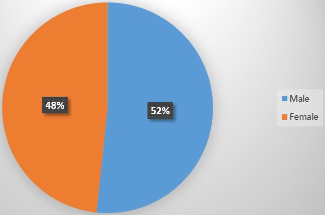
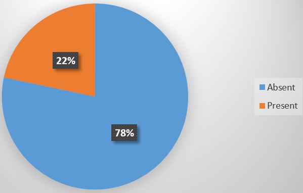
Results: Among the 60 newborns studied, the mean age of neonates was 12.45 ± 7.16 days with a male-to-female ratio of 1.1:1. Frequency of Meningitis in babies with late-onset sepsis was 21.7% (13 out of 60).

Conclusion: Meningitis is commonly associated with late-onset neonatal sepsis hence LP should be done as standard protocol in such neonates. This study demonstrated that the frequency of meningitis in late-onset neonatal sepsis was 21.7% (13/60).

Keywords: Meningitis, Late Onset, Neonatal Sepsis

Corresponding Author How to Cite this Article To Browse
Lubna Ahmed, Junior Consultant, Paediatrics, Sarkari Karmachari Hospital, Dhaka, , Bangladesh.
Email:
Ahmed L, Banik SK, Shahidullah S, Nesa V, Ema N, Arefin MN, Frequency of Meningitis in Late Onset Neonatal Sepsis-A cross-sectional descriptive study. Pediatric Rev Int J Pediatr Res. 2024;11(3):22-28.
Available From
https://pediatrics.medresearch.in/index.php/ijpr/article/view/770

Manuscript Received Review Round 1 Review Round 2 Review Round 3 Accepted
2024-08-07 2024-08-13 2024-08-19 2024-08-25 2024-08-31
Conflict of Interest Funding Ethical Approval Plagiarism X-checker Note
None Nil Yes 13.32

© 2024by Ahmed L, Banik SK, Shahidullah S, Nesa V, Ema N, Arefin MNand Published by Siddharth Health Research and Social Welfare Society. This is an Open Access article licensed under a Creative Commons Attribution 4.0 International License https://creativecommons.org/licenses/by/4.0/ unported [CC BY 4.0].

Introduction

Neonatal sepsis is one of the direct causes of neonatal mortality and is responsible for approximately 36% of the four million neonatal deaths that occur annually [1]. Neonatal sepsis can be categorized as early onset and late onset depending on whether the onset of symptoms is before 72 hours of life (Early onset) or after 72 hours of life (late onset). Late-onset sepsis (LONS) is caused by the organism present in the external environment of the home or the hospital [2]. The signs and symptoms of neonatal sepsis consist of fever or hypothermia, cyanosis, respiratory difficulties, apnea, reluctant to feed, lethargy, or irritability, hypotonia, fits, bulging fontanelle, bleeding problems, abdominal distension, unexplained jaundice, or more importantly “not looking well”[3]. Late-onset neonatal sepsis occurs in 0.1% of all newborns and up to 25% of very low birth weight infants (birth weight <1500 gm)[4]. Meningitis is the common presentation of LONS and it results in serious neurological sequelae and impairment [5]. The initial signs of neonatal meningitis, such as temperature instability, lethargy, apnea and bradycardia are often subtle and nonspecific and may occur as a result of other non-infectious etiologies [6]. Classic meningitic signs such as bulging fontanelle and seizures are usually found later in the course of illness [7]. It is almost impossible to distinguish sepsis from meningitis in the neonate clinically. However cerebrospinal fluid (CSF) positive for pathogenic bacteria indicates meningitis [8].In early-onset neonatal sepsis, lumbar puncture is indicated in the presence of a positive blood culture or if the clinical picture is consistent with septicaemia. It is not indicated if antibiotics have been started solely due to the presence of risk factors. In situations of late-onset sepsis, LP should be done in all infants before starting antibiotics. However, in clinically sick neonates lumbar puncture should be performed once the clinical condition stabilizes [9]. A total of 15-55% of patients who have meningitis (positive CSF culture) have negative blood cultures [10]. The most reliable way to diagnose meningitis is by obtaining a CSF specimen for analysis via a lumbar puncture (LP). If the CSF specimen is obtained before the initiation of antibiotics, the causative pathogen can be identified and appropriate antibiotic therapy can be determined[11].

Materials and Methods

Type of Study: Cross-sectional descriptive study.

Place of study: Department of Neonatology, SSMCMH, Mitford, Dhaka, Bangladesh.

Study Period: Twelve Months (November 2019 to October 2020).

Study Population: All the patients with LONS during the study were considered as the study population.

Study sample: Neonates with Late Onset Neonatal Sepsis.

Sample size calculation: Estimation of sample size by following formula:

n=(z²pq )/(d[12]

n = 149.23 ≈149 (estimated sample size)

Sample size: The study was carried out with a sample size of 60 due to the COVID-19 pandemic situation.

Selection criteria

Inclusion criteria: Babies with signs and symptoms of sepsis after 72 hours of life with the following criteria:

1. Apnea, tachypnea, respiratory distress, cyanosis
2. Bradycardia, tachycardia.
3. Hypotonia, seizures.
4. Poor skin colour, poor perfusion.
5. Irritability, lethargy, poor feeding.
6. Abdominal distension, hepatomegaly, splenomegaly.
7. Hypothermia, hyperthermia.

Exclusion criteria: Babies with the following signs and symptoms:

1. Congenital infection.
2. Early neonatal sepsis.
3. Perinatal asphyxia.
4. Congenital anomalies.
5. Intracranial haemorrhage.
6. Traumatic lumbar puncture

Study procedure: This study was conducted over 12 months (from November 2019 to October 2020) in the NICU, department of Neonatology of SSMC& MH, a tertiary care Hospital of Dhaka after approval by the Ethical committee.


A written informed consent was taken from parents. In each neonate with clinical features of sepsis, a detailed history and clinical examination were done. All babies with a provisional diagnosis of sepsis underwent complete blood count, CRP, blood C/S, chest x-ray and lumbar puncture.

Lumbar puncture was performed with appropriate aseptic precaution and by inserting a needle between the fourth and fifth lumber vertebrae (L4- L5). Routine examination of CSF includes physical, cytological, biochemical and microbiological examination. CSF was analysed as per standard laboratory methods.

The appearance and pressure of CSF were noted in physical examination, for cytological examination Neubauer counting chamber was used. For glucose estimation oxidase method was used (Dimension 100), and for protein estimation biuret method was used (Dimension-100). For gm staining and C/S, the CSF was sent to the microbiology laboratory.

Statistical Methods:

After collection, data were entered into a personal computer for analysis, and plotting and were presented in graphs and tables. Computer-based SPSS (Statistical Package for Social Science) version 23.0 was used. Quantitative data were expressed as the mean ± standard deviation and categorical data were presented as frequency and percentage. P< 0.05 was considered as statistically significant.

Results

During this period total of 60 newborns with LONs were included in this study. The data were arranged according to objectives set for the study and the results were presented in Tables and Graphs.

Demographic variables of mothers, among the 60 mothers, 51 (85.0%) mothers had age group ≤29 years, 35 (58.3%) were primipara, 46 (76.7%) took regular antenatal checkups and 30 (50.0%) belonged to low socioeconomic condition.

Distribution of delivery variables of mothers, out of 60 mothers, 52 (86.7%) had a gestational age between 37-42 weeks, 29 (48.3%) had undergone caesarean section, 41 (68.3%) had hospital delivery, premature rupture of membrane (PROM) and prolong labour were present in 3 (5%) and 4 (6.7%) cases respectively.

Table 1: Distribution of the neonates according to demographic variables (n=60)

Neonatesdemographic variablesFrequencyPercent(%)
Age(days)
≤103050.0
11-202135.0
21-28915.0
Mean±SD(Min-Max)12.45± 7.16(4-28)
Gender
Male3151.7
Female2948.3
Birthweight(gm)
<2500gm1220.0
≥2500gm4880.0
Mean±SD(Min-Max)2674.17 ± 464.82 (1200-3800)

Demographic variables of neonates, among 60 neonates, 30 (50.0%) neonates had an age ≤10 days, 21 (35%) had an age group lying between 11-20 days, male preponderance 31 (51.7%) and 48 (80.0%) neonates having ≥2500 gm birth weight (Table-I).

Among the study population 31(51.7%) neonates male and 29(48.3%) female as shown in figure-1

pediatric_770_01.JPG
Figure 1:
Pie chart showing gender distribution of the neonates.

Table 2: Distribution of the neonates according to the number of clinical findings (n=60)

Number of clinical findingsFrequencyPercent(%)
Single4066.7
Doubleormore2033.3
Total60100.0

Among 60 neonates, clinical variables were respiratory problems, fever, poor feeding or convulsion and 20 (33.3%) had double or more clinical findings (Table II).


Among the 60 neonates, the diagram shows that 50% of neonates have poor feeding, 35% have a fever, 28.3% have a respiratory problem and 21.7% have a convulsion as shown in figure-2.

pediatric_770_02.JPG
Figure 2:
Bar diagram showing the frequency of clinical findings among the neonates (n=60)

Table 3: Laboratory findings on blood analysis of neonates (n=60)

Investigation variablesMean ±SDMin-Max
CBG (mmol/L)3.95 ± 1.211.90-8.60
Hb (%)15.66 ± 2.2211.40-22.00
TC (pre/cumm)11202.67 ± 5807.743000.00-28300.00
Neutrophil (%)56.55 ± 15.2630.00-88.00
ANC6823.53 ± 4891.661290.00-22074.00
Platelet (per/cumm)254483.33 ± 570547.9232000.00-4550000.00

Among the 60 neonates, data regarding blood analysis shows septic screening, among them CBG 3.95 ± 1.21, Hb% 15.66 ± 2.22, total count of WBC 11202.67 ± 5807.74, neutrophil 56.55 ± 15.26, ANC (Absolute neutrophil count) 6823.53 ± 4891.66 and platelet count 254483.33 ± 570547.92 (Table III).

Table 4: Distribution of the neonates according to investigation variables (n=60)

Investigation variablesFrequencyPercent(%)
CRP
Positive4473.3
Negative1626.7
Blood CS
Growth46.7
No growth5693.3
X-ray
Normal5693.3
Abnormal46.7

CRP: C-reactive protein

Blood C/S: Blood culture and sensitivity

Abnormal X-ray means: patchy opacity or consolidation of the lung field

Table IV: Shows that different sepsis screening findings, among them CRP positive 44 (73.3%), Blood CS positive 4 (6.7%), chest x-ray findings present 4 (6.7%).

Table 5: Findings of CSF study among the neonates (n=60)

CSF studyMean±SDMin-Max
Cellcount(Total)39.93 ± 142.763.00-1000.00
Lymphocyte(%)93.60 ± 17.5910.00-100.00
Polymorphs(%)6.40 ± 17.59.00-90.00
Protein(mg/dl)183.71 ± 310.313.80-2300.00
Glucose(mmol/L)2.77 ± 1.270.50-8.40

Table V: Shows that CSF analysis of the neonates, Total cell count of CSF was 39.93 ± 142.76, lymphocyte 93.60 ± 17.59, polymorphs 6.40 ± 17.59, protein 183.71 ± 310.31 and sugar2.77 ± 1.27.

It was evident from the pie chart that meningitis was present in 13(21.7%) of LONS as shown in figure-3.

pediatric_770_03.JPG
Figure 3:
Frequency of Meningitis in neonates with Late Onset sepsis (n=60)

Discussion

Neonatal sepsis is one of the most common causes of neonatal morbidity and mortality. Near about 0.3-3% of neonates with sepsis do have meningitis but in the case of LONS, the frequency of meningitis is higher, even up to 30% Kaul et al[13]. This cross-sectional study was done in the Department of Neonatology in Sir Salimullah Medical College and Mitford Hospital,


Dhaka during the period from November 2019 to October 2020 for the detection of different CSF findings in late-onset neonatal sepsis and to determine the frequency of meningitis LONS. The overlapping clinical manifestations of septicemia and meningitis make it very difficult to differentiate a neonate with meningitis from one with septicemia alone as meningitis is associated with much more morbidity and mortality, it is always better to have a high suspicion for meningitis while treating neonates with septicemia [14].

In this study among 60 neonates, a male predominance was observed that is male to female ratio of 1.1:1. A Similar study conducted among neonates with sepsis by Das AK et al[9] 2020 found the male-to-female ratio of 2.2:1. All these studies support the result of our study and suggest the possibility of a sex-linked factor in host susceptibility and genetic susceptibility of the male patient to infection. In our part of the world, it might also be due to the patriarchal system of our society which gives more emphasis to the male child.

In this study, 20% (12/60) cases had birth weight less than 2500gm and 80% (48/60) babies had birth weight equal to more than 2500gm. In a study done in NIMCU and NICU of Kathmandu, Nepal in 2020, there were 29.6% of low birth weight (LBW) and 70.4% of appropriate weight babies. The lower incidence of LBW babies is due to the inclusion of only 13.8% (8 cases) of preterm babies. The symptoms with which the parents or caretakers presented in this study are almost comparable with the findings in the study by Das AK et al and Bhagat et al[9].

All these studies showed poor feeding, fever, respiratory problems and lethargy and/or convulsion to be the prominent features of presentation among newborns with sepsis which is comparable to our study. In this study, common features were poor feeding 50%, fever 35%, respiratory problems 28.3% and convulsion 21.7% of cases. In this study, CRP was positive in 73.3% of cases (44/60). In a study by Das AK et al [9] CRP was positive in 31.2% of cases (39/125). Among the 13 cases with abnormal CSF findings, CRP was positive in 11 cases (84.62%) and negative in 2 cases (15.38%). In a study by Ahmed A. Khattab et al. CRP was positive in 90% of cases. Among 20 cases with abnormal CSF findings,

CRP was positive in 11 cases (55%) and negative in nine cases (45%). In this study, blood culture was positive in 4 out of 60 cases, which account for 6.7% of total cases. In a similar study conducted in Nepal by Das et al [9] organisms were isolated in 21.6% of cases (27/125) of the collected blood samples. Another study was also conducted in Nepal by Shrestha NJ et al., organisms were isolated in 6.1% of the collected blood samples. Among 13 cases with abnormal CSF finding blood culture was positive in 2 cases (50.0%). In the present study isolated organism from blood culture was E.coli. The organism was sensitive to colistin, piperacillin/tazobactam, and tigecycline. In another study, E.coli was also isolated which is similar to our study but the isolated organism showed sensitivity to cloxacilin, amikacin, cotrimoxazole, cefixime, amoxicillin[9].

This sensitivity to organisms is different in our study probably due to demographic variables. In this study 21.7% (13 out of 60) neonates with late-onset neonatal sepsis were found to have abnormal CSF findings (greater than 10 leucocytes/cu mm in CSF) with the mean total leucocyte count was 39.93 ± 142.76 with a range of 3 to 1000, the mean protein level 183.71 ± 310.31, range 3.80 – 2300. The mean sugar level was 2.77 ± 1.27, range 0.50 to 8.40. In a study done by Das et al [9], it was found that 16% (20 out of 125) cases with LONS had abnormal CSF findings with cell count greater than 30 leucocytes/ cu mm in CSF.

In a study done by Saleem et al [15] it was found that 75 out of 198 patients (39.47%) of LONs had abnormal CSF, where the mean total leucocyte count was 24.36±22.27, the mean glucose level was 67.60±29.28 and mean protein was 127.14±77.35. In the present study, no organism was isolated from CSF. In a study done by Das AK et al [9] 0.8% (1 out of 125) presenting with LONS had positive CSF culture and the organism in the CSF culture was Streptococcus pyrogens. The organism was sensitive to cotrimoxazole and erythromycin.

Conclusion

This study demonstrated that a significant number of neonates with late-onset sepsis have co-existent neonatal meningitis (21.7%). Clinical features and blood analysis detect sepsis but cerebrospinal fluid findings lead diagnosis of neonatal meningitis.


Limitations

1. It is a single-centre study. So, results may not be representative of the whole population.
2. Small sample size due to COVID-19 pandemic situation during the study period.

Recommendations

1. A multicentre study with a large sample can be done to identify the prevalence of neonatal meningitis among newborns with late-onset neonatal sepsis.
2. Lumbar puncture and CSF examination is essential in all cases with Late Onset Neonatal Sepsis.

Conflicts of Interest: None

Financial Support and Sponsorship: Nil

Permission from Institutional research board: Yes

References

1. Shane AL, Stoll BJ. Neonatal sepsis: progress towards improved outcomes. J Infect. 2014 Jan;68 Suppl 1:S24-32. doi: 10.1016/j.jinf.2013.09.011. Epub 2013 Oct 18. PMID: 24140138 [Crossref][PubMed][Google Scholar]

2. Sankar MJ, Agarwal R, Deorari AK, Paul VK. Sepsis in the newborn. Indian J Pediatr. 2008 Mar;75(3):261-6. doi: 10.1007/s12098-008-0056-z. PMID: 18376095. [Crossref][PubMed][Google Scholar]

3. Shah BA, Padbury JF. Neonatal sepsis: an old problem with new insights. Virulence. 2014 Jan 1;5(1):170-8. doi: 10.4161/viru.26906. Epub 2013 Nov 1. PMID: 24185532; PMCID: PMC3916371. [Crossref][PubMed][Google Scholar]

4. Stoll BJ, Hansen N, Fanaroff AA, Wright LL, Carlo WA, Ehrenkranz RA, Lemons JA, Donovan EF, Stark AR, Tyson JE, Oh W, Bauer CR, Korones SB, Shankaran S, Laptook AR, Stevenson DK, Papile LA, Poole WK. Late-onset sepsis in very low birth weight neonates: the experience of the NICHD Neonatal Research Network. Pediatrics. 2002 Aug;110(2 Pt 1):285-91. doi: 10.1542/peds.110.2.285. PMID: 12165580. [Crossref][PubMed][Google Scholar]

5. Gupta N, Grover H, Bansal I, Hooda K, Sapire JM, Anand R, Kumar Y. Neonatal cranial sonography: ultrasound findings in neonatal meningitis-a pictorial review. Quant Imaging Med Surg. 2017 Feb;7(1):123-131. doi: 10.21037/qims.2017.02.01. PMID: 28275563; PMCID: PMC5337180 [Crossref][PubMed][Google Scholar]

6. Nizet, V. , Remington, J. S. , Klein, J. O., Wilson, C.B., Nizet, B. &Maldonaldo. 2010, Infectious Diseases of Fetus and Newborn Infant, 7thed, Philadelphia, PA, Saunders, pp. 243-248 [Crossref][PubMed][Google Scholar]

7. Krebs VL, Costa GA. Clinical outcome of neonatal bacterial meningitis according to birth weight. Arq Neuropsiquiatr. 2007 Dec;65(4B):1149-53. doi: 10.1590/s0004-282x2007000700011. PMID: 18345420.Kleion, J.O. & Remington, J.S. 2001, Infectious diseases of the fetus and newborn. 5th ed. Philadelphia, WB.Saunders [Crossref][PubMed][Google Scholar]

8. Das, AK, Mishra, D, Jha, N. K, Mishra, R. &Jha, S,. Role of Lumbar Puncture In Late Onset Neonatal Sepsis‟, J. Nepal PaediatrSoc, 2019:39(3):155-61 [Crossref][PubMed][Google Scholar]

9. Heath PT, Nik Yusoff NK, Baker CJ. Neonatal meningitis. Arch Dis Child Fetal Neonatal Ed. 2003 May;88(3):F173-8. doi: 10.1136/fn.88.3.f173. PMID: 12719388; PMCID: PMC1721548 [Crossref][PubMed][Google Scholar]

10. Remington, J. S. &Klein, J. O. 2001, Infectious diseases of the fetus and newborn. 5 thed, Philadelphia, WB Saunders [Crossref][PubMed][Google Scholar]

11. Riordan FA, Cant AJ. When to do a lumbar puncture. Arch Dis Child. 2002 Sep;87(3):235-7. doi: 10.1136/adc.87.3.235. PMID: 12193440; PMCID: PMC1719243 [Crossref][PubMed][Google Scholar]

12. Hoque, M. 2016, abc of research methodology and biostatistics. 2nd ed, Parveen Sultana Dhaka. Bangladesh. [Crossref][PubMed][Google Scholar]


13. Kaul V, Harish R, Ganjoo S, Mahajan B, Raina SK, Koul D. Importance of obtaining lumbar puncture in neonates with late onset septicemia a hospital based observational study from north-west India. J Clin Neonatol. 2013 Apr;2(2):83-7. doi: 10.4103/2249-4847.116407. PMID: 24049750; PMCID: PMC3775142 [Crossref][PubMed][Google Scholar]

14. Roshi, J. S. , Sheikh, Q. H. , Imran, A.G &Shabbir, A.W. 2015, „Incidence of Meningitis in Late Onset Neonatal Sepsis‟, J Microbial Immunol Infect, vol. 37, pp. 301-6 [Crossref][PubMed][Google Scholar]

15. Saleem, S. , Akbar, N. , Anwar, A. & Mehmood, R. Frequency of Meningitis inNeonatal Onset Sepsis‟, A.P.M.C, 2015:9(3):140-144 [Crossref][PubMed][Google Scholar]