E-ISSN:2349-3267
P-ISSN:2349-5499

Case Report

Myelopathy

Pediatric Review - International Journal of Pediatric Research

2020 Volume 7 Number 6 July-August
Publisherwww.medresearch.in

Compresetsive myelopathy presenting with paraparesis in pediatric age

Kotrashetti V. A.1, Baburao S V.2, Bainade K.3, Ravindran N S.4*, Vatkar A.5, Gupta S.6
DOI: https://doi.org/10.17511/ijpr.2020.i06.14

1 Kotrashetti V. A., Professor, Department of Pediatrics, DY Patil Hospital, Nerul, Maharashtra, India.

2 Vijay Baburao Sonawane, Associate professor, Department of Pediatrics, DY Patil Hospital, Nerul, Maharashtra, India.

3 Kapil Bainade, Assistant Professor, Department of Pediatrics, DY Patil Hospital, Nerul, Maharashtra, India.

4* Shweta Ravindran Nair, Senior Resident, Department of Pediatrics, DY Patil Hospital, Nerul, Maharashtra, India.

5 Amit Vatkar, Assistant Professor, Department of Pediatrics, DY Patil Hospital, Nerul, Maharashtra, India.

6 Samarth Gupta, Junior resident, Department of Pediatrics, DY Patil Hospital, Nerul, Maharashtra, India.

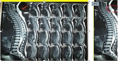
Compressive myelopathy is a common presentation of the adult age group secondary to degenerating etiologies. Trauma is a rare cause of compressive myelopathy, especially in the pediatric age group as the pediatric spine is more elastic than that of adults, especially below 8 years of age. Boys are more commonly injured as compared to girls. The present study reports a case of a six-year-old female patient of compressive myelopathy presenting with jerks with progressive paraparesis. MRI shows posterior subluxation of dens (C2) attached to C2 vertebral effacing anterior – subarachnoid space and indenting spinal cord.

Keywords: Spine injury, Dens dislocation, Atlanto-dental interval, Atlanto-axial fusion

Corresponding Author How to Cite this Article To Browse
Shweta Ravindran Nair, Senior Resident, Department of Pediatrics, DY Patil Hospital, Nerul, Maharashtra, India.
Email:
Kotrashetti V A, Sonawane VB, Bainade K, Nair S R, Vatkar A, Gupta S. Compresetsive myelopathy presenting with paraparesis in pediatric age. Pediatric Rev Int J Pediatr Res. 2020;7(6):313-316.
Available From
https://pediatrics.medresearch.in/index.php/ijpr/article/view/616

Introduction

About 1-5% of all spinal injuries occur in children [1-6]. Children below 8 years old have a relatively large and heavy head compared to the body which shifts the fulcrum of movement to the upper cervical spine (occiput to C2) making it more vulnerable to injury [7]. With growing age, fulcrum starts shifting to lower cervical in adults [8] This

explains the epidemiological finding that the majority of spinal injuries occur between C0 and C2 in young children whereas older children, like adults, have their injuries more commonly in the sub axial cervical spine [9]. The pediatric spine is more elastic than that of adults especially below 8 years of age [10] and it has been shown in neonates that the vertebral column could stretch by 2 inches without disruption whereas the spinal cord could only stretch by 0.25 inches [11]. Hence


Manuscript Received Review Round 1 Review Round 2 Review Round 3 Accepted
2020-08-04 2020-08-19 2020-08-23 2020-08-25
Conflict of Interest Funding Ethical Approval Plagiarism X-checker Note
No Nil Yes 4%

© 2020 by Kotrashetti V. A., Vijay Baburao Sonawane, Kapil Bainade, Shweta Ravindran Nair, Amit Vatkar, Samarth Gupta and Published by Siddharth Health Research and Social Welfare Society. This is an Open Access article licensed under a Creative Commons Attribution 4.0 International License https://creativecommons.org/licenses/by/4.0/ unported [CC BY 4.0].

trauma to the spine in young children can produce neural damage much earlier to musculoskeletal injury. The present study presents a case of a six-year-old female patient of compressive myelopathy with paraparesis.

Case Report

A 6-year-old female, developmentally normal, fully immunized was presented to the OPD with weakness of limbs. The child started complaining of pain in both lower limbs after which she had weakness in lower limbs. This continued over a month leading to difficulty in getting up and sitting on her own and walking without support. The patient also developed involuntary movements of the neck which began for one month and gradually increased in frequency. Initially, there was one episode in a day which gradually increased to 4-5 episodes in a day lasting for 5-10 seconds. Detailed history revealed a fall from the staircase 2 months back. There was the injury to the head with no intracranial bleeding, no ear nose, and throat bleeding, no chest or abdominal trauma. On examination, the patient was conscious, alert, and oriented to time, place, and person. Her vitals were normal. On systemic examination higher mental functions were normal along with normal cranial nerve examination.

The tone was normal in all four limbs; power in Upper limb (shoulder flexion, extension, abduction and adduction, elbow extension and flexion and wrist extension and flexion) was 3/5. Power in the lower limb (hip extension, hip abduction) was 2/5 whereas, in hip flexion, adduction and knee extension and flexion and foot dorsiflexion were 3/5. Deep tendon reflexes were brisk and ankle clonus was present. Sensory examination touch, pain, temperature sensations are normal. There were no signs of cerebellar involvement and meningeal irritation. Routine and metabolic workup was normal. The child was initiated on Levetiracetam after which seizures improved. MRI Brain and Spine (Figure 1) screening is done as it is the investigation of choice [12].

EEG (Figure 2) showed intermittent sharp wave activity of the alpha rhythm in the range of 8-13Hz. It revealed posterior subluxation of dens causing significant cord compression at the level of the dens. Residual spinal cord diameters at this level measure 6.2mm. There is a high T2 signal noted within the cord at these levels suggestive of compressive myelopathy.

A dedicated MRI Cranio-cervical Junction was done which suggested posterior subluxation of dens (C2) attached to C2 vertebral effacing anterior – subarachnoid space and indenting spinal cord. The MRI also showed increased atlanto-dental interval measuring 6.3 mm maximum anteroposterior dimension. Hyperintense signal intensity is also noted involving the spinal cord at this level measuring approximately 2 cm in length suggestive of compressive myelopathy. CT Angiography revealed the caliber of the right vertebral artery is significantly smaller than the left artery. Injection Dexamethasone was started for the reduction of inflammation in the spine by a pediatric neurologist. For further management of cervical fusion procedure (occipito-cervical fusion) was planned.

pedia_616_01.jpg

Fig-1: MRI showing cord compression at the C1-C2 level.

pedia_616_02.jpg

Fig-2: EEG showing intermittent sharp wave activity of alpha rhythm.

Discussion

Our patient was presented to the OPD with weakness of limbs. On detailed history, fall was revealed following which the child started showing involuntary movements of the neck. Differentials of acute paralysis were considered. Since the child did not have ascending paralysis and was completely immunized for age with brisk reflexes, intact sensations, with no evidence of an acute injury differential of Guillain barre syndrome [13], acute poliomyelitis, transverse myelitis were ruled out.


There is evidence from studies that traumatic compressive myelopathy is less common in the pediatric age group and more common in adults [1]. But after routine investigation ruling out common etiologies like degenerative causes, infection, congenital causes lead to suspicion of traumatic compressive myelopathy.

This condition is usually detected radiographically in the evaluation of persistent problems after cervical trauma or symptoms of spinal cord dysfunction. This confirmed our diagnosis of Cervical Compressive Myelopathy at C-V junction secondary to trauma. Inflammation was also present for which Dexamethasone injection was started. To reduce the compression till surgical intervention a cervical collar was recommended.

Management involves a form of atlantoaxial fixation and fusion if atlantoaxial dislocation can be reduced by traction. It is a safe and predictable procedure [14]. Occipital-cervical fusion may be required if irreducible atlantoaxial dislocation exists, or if the spinal canal sagittal diameter is less than 13 mm and C1 laminoplasty should be performed. So, the surgical intervention was planned for the patient in coordination with a pediatric neurosurgeon.

Conclusion

Atlantoaxial instability increases the risk of developing myelopathy and acute cervical cord injury after minor trauma in patients. Presentation of a child with paraparesis and history of trauma should alert the physician for a probable diagnosis of myelopathy though rare. Fixation and fusion should be performed to prevent future myelopathy and to improve neurological symptoms due to acute traumatic cervical cord injury in patients with atlantoaxial instability.

Reference

  1. Nouri A, Tetreault L, Singh A, Karadimas SK, Fehlings MG. Degenerative cervical myelopathy- epidemiology, genetics, and pathogenesis. Spine. 2015;40(12)E675-E693.
  2. Kewalramani LS, Tori JA. Spinal cord trauma in children, Neurologic patterns, radiologic features, and pathomechanics of injury. Spine. 1980;5(1)11-18.
  1. Anderson JM, Schutt AH. Spinal injury in children- a review of 156 cases seen from 1950 through 1978. InMayo Clinic Proceedings. 1980;55(8)499-504.
  2. Bracken MB, Freeman DH Jr, Hellenbrand K. Incidence of acute traumatic hospitalized spinal cord injury in the United States, 1970-1977. Am J Epidemiol. 1981;113(6)615-622.
  3. Hamilton MG, Mylks ST. Pediatric spinal injury- review of 174 hospital admissions. J Neurosurg. 1992;77(5)700-704.
  4. d’Amato C. Pediatric spinal trauma- injuries in very young children. Clin Orthopaed Rel Res. 2005;432;34-40.
  5. Birney TJ, Hanley Jr EN. Traumatic cervical spine injuries in childhood and adolescence. Spine. 1989;14(12)1277-1282.
  6. Townsend EH, Rowe ML. Mobility of the upper cervical spine in health and disease. Pediatr. 1952;10(5)567-574.
  7. Hadley MN, Zabramski JM, Browner CM, Rekate H, Sonntag VK. Pediatric spinal trauma- review of 122 cases of spinal cord and vertebral column injuries. J Neurosurg. 1988;68(1)18-24.
  8. Herkowitz H N, Rothman R H. Subacute instability of the cervical spine. Spine. 1984;9(4)348-357.
  9. Leventhal H R. Birth injuries of the spinal cord. J Pediatr. 1960;56(4)447-453.
  10. Flynn J M, Closkey R F, Mahboubi S, Dormans J P. Role of magnetic resonance imaging in the assessment of pediatric cervical spine injuries. J Pediatr Orthop. 2002;22(5)573-577.

  1. Alter M. The epidemiology of Guillain-Barr syndrome. Ann Neurol. 1990;27(S1)S7-S12.
  2. Smith MD, Phillips WA, Hensinger RN. Fusion of the upper cervical spine in children and adolescents- an analysis of 17 patients. Spine. 1991;16(7)695-701.