E-ISSN:2349-3267
P-ISSN:2349-5499

Case Report

Colonic

Pediatric Review - International Journal of Pediatric Research

2020 Volume 7 Number 8 November-December
Publisherwww.medresearch.in

A novel RET gene mutation in a neonate with total colonic aganglionosis and renal agenesis: a case report

Teja Goud M R.1, Kumar K.2, Banu S.3*
DOI: https://doi.org/10.17511/ijpr.2020.i08.08

1 Ravi Teja Goud M, DNB (Pediatric), Department of Neonatology, Ankura Hospital for Women and Children, Hyderabad, Telangana, India.

2 K. Lalatendu Kumar, Consultant, Pediatric Surgeon and Pediatric Urologist, Ankura Hospital for Women and Children, Hyderabad, Telangana, India.

3* S. Nasreen Banu, Consultant Neonatologist, Department of Neonatology, Ankura Hospital for Women and Children, Hyderabad, Telangana, India.

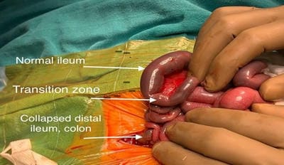
Hirschsprung’s disease (HSCR) is functional lower intestinal obstruction, due to the congenital absence of the intramural plexuses of ganglion cells in the distal bowel. Total colonic aganglionosis (TCA) is a rare and severe form of HSCR and accounts for 5-10% of all the diagnosed cases of HSCR. TCA is a diagnostic and therapeutic challenge as clinical and radiological findings are not pathognomonic. The RET gene signaling system is generally acknowledged as being the most important in TCA pathogenesis, with RET gene variations being present in 70% of cases. The present study is reporting a case of TCA and right renal agenesis with a novel mutation in the RET gene.

Keywords: Hirschsprung’s disease (HSCR), Total colonic aganglionosis (TCA), REarranged during Transfection gene (RET)

Corresponding Author How to Cite this Article To Browse
S. Nasreen Banu, Consultant Neonatologist, Department of Neonatology, Ankura Hospital for Women and Children, Hyderabad, Telangana, India.
Email:
Teja Goud MR, Kumar KL, Banu SN. A novel RET gene mutation in a neonate with total colonic aganglionosis and renal agenesis: a case report. Pediatric Rev Int J Pediatr Res. 2020;7(8):437-441.
Available From
https://pediatrics.medresearch.in/index.php/ijpr/article/view/635

Manuscript Received Review Round 1 Review Round 2 Review Round 3 Accepted
2020-10-30 2020-11-01 2020-11-20 2020-12-09
Conflict of Interest Funding Ethical Approval Plagiarism X-checker Note
No Nil Yes 6%

© 2020 by Ravi Teja Goud M, K. Lalatendu Kumar, S. Nasreen Banu and Published by Siddharth Health Research and Social Welfare Society. This is an Open Access article licensed under a Creative Commons Attribution 4.0 International License https://creativecommons.org/licenses/by/4.0/ unported [CC BY 4.0].

Introduction

Hirschsprung’s disease (HSCR) is the main genetic cause of functional intestinal obstruction with an incidence of 1/5000 live births. Total colonic aganglionosis (TCA) is a rare and severe form of HSCR with the incidence being 1 in 5,00,000 [1]. TCA accounts for 5-10% of all the diagnosed cases of HSCR [2]. The genes implicated in the pathogenesis of HSCR have related the two main gene susceptibility pathways identified, the RE arranged during Transfection (RET) signaling cascade and the endothelin B receptor-related pathways.

At least 12 genes have been identified to date and are continually being added to [3]. The RET gene signaling system is the most important mechanism in TCA pathogenesis and accounts for about 70% of cases [4]. Genetic diagnosis helps in genetic counseling and prenatal diagnosis and hence this is an attempt to report a case of TCA with a novel mutation in the RET gene.

Case Report

A 4-day old neonate was brought to the emergency department with complaints of bilious vomiting, abdominal distension, and non-passage of meconium since birth. The baby of S, a full term, 39+4 week male neonate born to G3P1L1A1 mother with 3rd-degree consanguinity, by lower segment cesarean section. The baby had a normal transition with a birth weight of 2800 grams. No obvious external congenital anomalies were seen. The baby was started on exclusive breastfeeds.

On day 2 of life baby developed bilious vomiting and abdominal distension. Antenatal ultrasonography showed hypoplastic nasal bone and absent or atrophic right kidney for which amniocentesis was done at 25 weeks of gestation to evaluate for chromosomal aneuploidy and it was normal. Chromosomal microarray showed deletion in sex chromosomes (implies causing infertility).

The baby was kept nil by mouth and started on total parenteral nutrition(TPN). The X-ray showed dilated bowel loops. USG abdomen showed non visualized right kidney and ureterocele. The lower GI contrast study was inconclusive (did not show the transition zone). Given persistent bilious vomiting, worsening abdominal distension, and non-passage of meconium despite rectal stimulation, a pediatric surgeon was involved.

Exploratory laparotomy and ileostomy were done on day 6 of life. Intra-operative findings included dilated proximal small bowel loops with a doubtful transition zone at the distal ileum, 20 cm proximal to the ileocecal valve, and collapsed distal bowel (Figure1). The possibility of TCA was considered and multiple seromuscular biopsies from the rectum, transverse colon, appendix, transition zone (distal ileum), and stoma site were sent for histopathology.

Post-operatively baby was continued on TPN. As the stoma was not functioning elemental formula feeds couldn’t be established even by postoperative day 9. Biopsy showed the absence of ganglion cells and absence of neural hypertrophy in all sections including that from the stoma site, which confirmed TCA. Given aganglionosis at the stoma site, and no improvement postoperatively, a plan was made to resite the stoma.

pedia_635_01.jpg

Fig-1: Intraoperative picture showing transition zone approximately 20cm proximal to the ileocecal valve and collapsed distal bowel.

As a part of further evaluation, clinical exome sequencing was sent, which showed a mutation in the RET gene which is a known gene involved in TCA. A homozygous missense variation in exon 19 of the RET gene (chr10:g.43126712C>A; Depth: 123x) that results in the amino acid substitution of Lysine for Asparagine at codon 1059 (p.Asn1059Lys) was detected. This variant has not been previously reported in the RET gene in association with HSCR.

Parents were counseled about the need for the laparotomy and resiting of the stoma and the possibility of long term complications like short bowel syndrome and mortality. Parents decided to continue further treatment at a government hospital due to personal reasons. At the government, hospital the baby was treated conservatively and succumbed on day 45 of life.


Discussion

HSCR is a congenital malformation of the gut characterized by the absence of parasympathetic intrinsic ganglion cells in the submucosal and myenteric plexuses in the distal bowel [5].

It is due to the premature arrest of the craniocaudal migration of vagal neural crest cells in the hindgut between the fifth and twelfth week of gestation to form the enteric nervous system (ENS) and is therefore regarded as a neurocristopathy. Depending on the extent of involvement it is classified into ultra-short-segment, short segment(S-HSCR), and long segment (L-HSCR) [6].

The long-segment disease can be further divided into long-segment colonic aganglionosis, total colonic aganglionosis (TCA), and total colonic and small bowel aganglionosis (TCSA). The latter when it involves a very-long-segment it is called Zuelzer syndrome and total bowel aganglionosis is also reported [7]. In TCA, aganglionosis extends from the anus to at least the ileocecal valve, but not 50 cm proximal to it [8].

TCA is a rare and severe form of HSCR [1]. TCA is much more common in female individuals, and the 4:1 male predominance of S-HSCR decreases to 1.1:1 or even 0.8:1 for TCA [9]. HSCR though considered as multifactorial, genetic factors play a major role in its pathogenesis. The recurrence risk of HSCR is approximately 200 times higher in the affected families and higher in cases of longer aganglionic segments and TCA, presumably due to increased gene penetrance [10].

The genes implicated in the pathogenesis of HSCR are related to one of the two main gene susceptibility pathways identified (the RE arranged during Transfection signaling cascade [RET, glial cell line-derived neurotrophic factor (GDNF), glial cell line-derived neurotrophic factor family receptor alpha (GFR), neurturin (NTN)] and the endothelin B receptor-related pathways [endothelin receptor type B (EDNRB), endothelin 3 (EDN3), endothelin converting enzyme 1 (ECE-1), PHOX2, and SRY-box containing gene (SOX10)]).

Other identified genes are mostly related to specific syndromes, and their pathogenetic connection to HSCR is not as yet fully established. Identification of gene assists in genetic counseling, particularly in potential familial recurrences.

The RET gene signaling system is generally acknowledged as being the most important in TCA pathogenesis, with RET gene variations being present in 70% of cases [4]. Previously reported RET gene mutations associated with TCA, typically have deletions or frameshifts, such as N302EfsX53, K549_G550delD, V636fsX1D, K549_G550delD, and V145GD [11]. The index case was diagnosed to have homozygous missense mutation p.Asn1059Lys in the RET gene. Analysis of the specific mechanisms of pathogenesis involving the p.Asn1059Lys mutation is necessary.

Due to its rarity, TCA is still a diagnostic and therapeutic challenge. Clinical and radiologic findings can be useful in diagnosis, but they are not pathognomonic. N-Fekete et al, reported that there is a high rate of TCA cases that passed meconium in the first 24 hours of life [12]. A high index of suspicion is required for the early diagnosis of TCA because the usual symptoms like the delayed passage of meconium, abdominal distension, and bilious vomiting are infrequently seen.

However, the index case presented with the typical complaints of HSCR.Per rectal examination may not result in explosive passage of feces as seen in classic HSCR, due to a less abrupt transition from normal to the aganglionic segment. The radiographic picture is often inconclusive. Abdominal X-rays may show features of intestinal obstruction and a variable degree of dilatation of bowel loops above the aganglionic segment as seen in the index case. Fondalli et al, concluded that microcolon with reflux into mega ileum appears to be the most suggestive finding for TCA [13], which is not found in our case.

Tsuji et al, reported that 25% of their patients had associated anomalies in the form of cardiovascular, gastro-intestinal, or urogenital anomalies [14]. A study in mice showed a homozygous mutation in the RET gene which was associated with renal agenesis along with L-HSCR[15]. Index case has right renal agenesis and ureterocele along with TCA. A similar case of TCA with right renal agenesis and oligomeganephroma (OMN) has been reported by Sugimoto et al, found to have a heterozygous point mutation p.S811F in exon 14 of RET gene, changing serine to phenylalanine [16].

Recognition of TCA is hard during laparotomy as false transition zones can be seen [2]. The transition zone is often long in TCA and interpretation of frozen sections can be difficult.


The definitive diagnosis is made following suction / open biopsy of the rectum, colon, and ileum. If this condition is suspected pre-operatively, frozen section biopsies should be ideally done to assess the level of aganglionosis to determine the level of diverting stoma, as many times clear transition zone could not be visible in TCA.

This may lead to wrong placement of stoma in an aganglionic segment, which is a very common operative error [17] as seen in the index case. The absence of ganglion cells in submucosal and myenteric plexuses confirms the diagnosis of HSCR. Additionally, the presence of hypertrophic nerve fibers may aid in the diagnosis, but it is not so in TCA, similar to the index case.

The mortality rate is high and most of them require long-term parenteral nutrition. Multiple operative procedures like Swenson, Martin, Duhamel, Martin modification of Duhamel, Kimura, endorectal pull through, Rehbein, and direct ileorectal anastomosis are mentioned for TCA, but there is no consensus regarding the superiority of one over the other [18].

Conclusion

Affected families have a 200 times higher risk of HSCR recurrence and are even higher with TCA. Here the current study reports a neonate with TCA and concomitant right renal agenesis and ureterocele. A novel homozygous missense mutation, p.Asn1059Lys in exon 19 of the RET gene was identified. This case report may help to establish the prenatal diagnosis and appropriate genetic counseling.

Reference

  1. Ikeda K, Goto S. Diagnosis and treatment of Hirschsprung's disease in Japan- An analysis of 1628 patients. Ann Surg. 1984;199(4)400-405.
  2. Dykes EH, Guiney EJ. Total colonic aganglionosis. J Pediatr Gastroenterol Nutr. 1989;8(1)129-132.
  3. Moore SW. Hirschsprung disease- Current perspectives. Open Access Surg. 2016;9;39-50.
  1. Moore SW. Total colonic aganglionosis in Hirschsprung disease. Semin Pediatr Surg. 2012;21(4)302-309.
  2. Whitehouse FR, Kernohan JW. Myenteric plexus in congenital megacolon- study of 11 cases. Arch Intern Med (Chic). 1948;82(1)75-111.
  3. Kaiser G, Bettex M. Disorders and congenital ma lformations associated with Hirschsprung’s dise ase, In- Holschneider AM, ed, Hirschsprung’s Dis ease. Stuttgart-Hipokrates-Verlag. 1982;49-53.
  4. ZUELZER WW, WILSON JL. Functional intestinal obstruction on a congenital neurogenic basis in infancy. Am J Dis Child. 1948;75(1)40-64.
  5. Hoehner JC, Ein SH, Shandling B, Kim PC. Long-term morbidity in total colonic aganglionosis. J Pediatr Surg. 1998;33(7)961-965.
  6. Louw JH. Total colonic aganglionosis. Can J Surg. 1978;21(5)397-404, 409.
  7. Amiel J, Lyonnet S. Hirschsprung disease, associated syndromes, and genetics- a review. J Med Genet. 2001;38(11)729-739.
  8. So MT, Leon TY, Cheng G, Tang CS, Miao XP, Cornes BK, et al. RET mutational spectrum in Hirschsprung disease- evaluation of 601 Chinese patients. PLoS One. 2011;6(12)e28986.
  9. N-Fékété C, Ricour C, Martelli H, Jacob SL, Pellerin D. Total colonic aganglionosis (with or without ileal involvement)- a review of 27 cases. J Pediatr Surg. 1986;21(3)251-254.
  10. Fondelli P, Martucciello G, Taccone A, Dodero P. Quadro radiologicodell agangliatotale del colon [The radiologic picture of total colonic aganglionosis]. Radiol Med. 1989;77(3)207-10.

  1. Tsuji H, Spitz L, Kiely EM, Drake DP, Pierro A. Management and long-term follow-up of infants with total colonic aganglionosis. J Pediatr Surg. 1999;34(1)158-161.
  2. Schuchardt A, D'Agati V, Larsson-Blomberg L, Costantini F, Pachnis V. Defects in the kidney and enteric nervous system of mice lacking the tyrosine kinase receptor Ret. Nature. 1994;367(6461)380-383.
  1. Sugimoto K, Miyazawa T, Nishi H, Miyazaki K, Enya T, Okada M, et al. Heterozygous p-S811F RET gene mutation associated with renal agenesis, oligomeganephronia and total colonic aganglionosis- a case report. BMC Nephrol. 2016;17(1)146.
  2. Yk S, P R, N T. Perils of total colonic aganglionosis presenting in neonatal age. J Neonatal Surg. 2014;3(3)28.
  3. Chandelia S, Nangia S, Saili A. Total colonic aganglionosis- a diagnostic intricacy. Indian J Pediatr. 2013;80(9)789-790.