E-ISSN:2349-3267
P-ISSN:2349-5499

Research Article

Blood

Pediatric Review - International Journal of Pediatric Research

2020 Volume 7 Number 8 November-December
Publisherwww.medresearch.in

Thyroid profile in patients of thalassemia with multiple blood transfusions and high serum ferritin: a cross-sectional study

Khandelwal R.1, Gundluru M.2*, Mehta K. L.3
DOI: https://doi.org/10.17511/ijpr.2020.i08.02

1 Rohit Khandelwal, Associate Professor, Department of Paediatrics, Vydehi Institute of Medical Sciences and Research Centre, Bangalore, Karnataka, India.

2* Muralidhar Gundluru, Assistant Professor, Department of Paediatrics, Vydehi Institute of Medical Sciences and Research Centre, Bangalore, Karnataka, India.

3 Leeni Mehta K., Consultant Physician, Apollo Group of Hospitals, Bangalore, Karnataka, India.

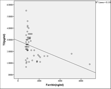
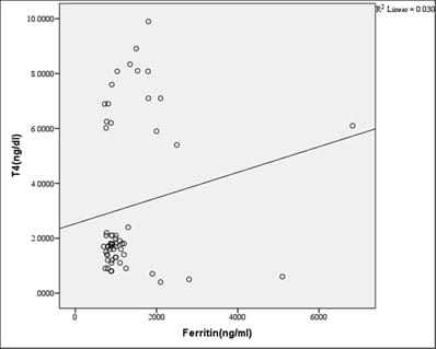
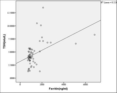
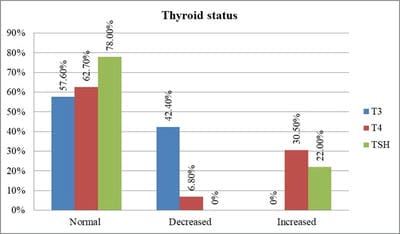
Introduction: Beta-thalassemia major patients undergo regular blood transfusion resulting in growth faltering and various endocrine problems including thyroid dysfunction due to iron overload in the body. This study was conducted to determine the frequency of thyroid dysfunction in children presenting with Beta-thalassemia major on regular blood transfusions. Materials and methods: Sixty children were included with proven beta-thalassemia major who reported to the Department of Pediatrics, VIMS, and RC, Bangalore. Inclusion criteria: 1.Children 4 to 18 years age group .2.The child received transfusions for more than 2 years. 3.Children with serum ferritin level >700. Results: In this study, four patients(6.8%) had overt hypothyroidism, eight patients(13.6%) had subclinical hypothyroidism and 47 patients(79.7%) had euthyroid status. There was a positive correlation between Ferritin and T4, TSH levels. i.e., with an increase in Ferritin level, there was an increase in T4, TSH levels, and vice versa. However, the correlation was significant with TSH. There was a significant negative correlation between Ferritin and T3 levels. i.e with an increase in Ferritin level, there was a decrease in T3 levels and vice versa. Conclusion: Thyroid dysfunction can exist in thalassemia patients on multiple transfusions and chelation therapy with high serum ferritin levels. Detection of hypothyroidism is important as inexpensive oral replacement therapy is readily available. Hence regular screening of beta-thalassemia major patients for Serum T3, Serum T4, Serum TSH for early detection and timely treatment could improve the life expectancy and quality of life of these patients.

Keywords: Blood transfusion, Hypothyroidism, Serum Ferritin, Serum T3, Serum T4, Serum TSH, Thalassemia

Corresponding Author How to Cite this Article To Browse
Muralidhar Gundluru, Assistant Professor, Department of Paediatrics, Vydehi Institute of Medical Sciences and Research Centre, Bangalore, Karnataka, India.
Email:
Khandelwal R, Gundluru M, Mehta KL. Thyroid profile in patients of thalassemia with multiple blood transfusions and high serum ferritin: a cross-sectional study. Pediatric Rev Int J Pediatr Res. 2020;7(8):401-408.
Available From
https://pediatrics.medresearch.in/index.php/ijpr/article/view/646

Manuscript Received Review Round 1 Review Round 2 Review Round 3 Accepted
2020-12-15 2020-12-21 2020-12-25 2020-12-28
Conflict of Interest Funding Ethical Approval Plagiarism X-checker Note
No Nil Yes 7%

© 2020 by Rohit Khandelwal, Muralidhar Gundluru, Leeni Mehta K. and Published by Siddharth Health Research and Social Welfare Society. This is an Open Access article licensed under a Creative Commons Attribution 4.0 International License https://creativecommons.org/licenses/by/4.0/ unported [CC BY 4.0].

Introduction

Beta-thalassemia is a group of hereditary blood disorders characterized by anomalies in the synthesis of the β chains of hemoglobin resulting in variable phenotypes ranging from severe anemia to clinically asymptomatic individuals [1].

The total annual incidence of symptomatic individuals is estimated at 1 in 100,000 throughout the world and 1 in 10,000 people in the European Union [2]. Earlier studies have shown that the overall prevalence of β-thalassemia is 3–4 % with an estimate of around 8,000 to 10,000 new births with the major disease each year [3,4].

The homozygous state results in severe anemia, which needs a regular blood transfusion, The combination of transfusion and chelation therapy has dramatically extended the life expectancy of thalassemic patients who can now survive into their fourth and fifth decades of life [5,6]. If a regular transfusion program that maintains a minimum Hb concentration of 9.5 to 10.5 g/dL is initiated, growth and development tend to be normal up to 10 to 12 years [7].

Regular transfusion therapy leads to iron overload related complications including endocrine complication (growth retardation, failure of sexual maturation, diabetes mellitus, and insufficiency of the parathyroid, thyroid, pituitary, and less commonly, adrenal glands), dilated myocardiopathy, liver fibrosis, and cirrhosis) [8].

These complications are due to the toxic effects of the iron that gets deposited in the endocrine glands. Thyroid hormone is essential for the development and maintenance of normal function of CNS including brain development and its deficiency leads to mental retardation.

So regular follow-up for early detection and timely treatment of such complications could improve the quality of life of these patients. Very few studies comparing the frequency of thyroid dysfunction in children presenting with Beta-thalassemia major have been done. Hence this study was conducted to know the thyroid function status and timely treatment can be started along with the primary management.

Objective of study

To determine the frequency of thyroid dysfunction in children presenting with Beta-thalassemia major.

Materials and methods

Source of data: Children in the age group 5 years-18 years who reported to OPD and IPD of the Department of Pediatrics in VIMS and RC from Jan 2017 to June 2018. Assuming a prevalence of 2-14%, a sample size of 60 Beta thalassemia major patients were included in this study.

Inclusion criteria:

1.Children 4 to 18 years age group with proven Beta thalassemia major.

2.Child received blood transfusions for more than 2 years.

3.Children with serum ferritin level >700(ng/ml).

Exclusion criteria:

1.Children with thalassemia minor or intermedia.

2.Children less than 4 years of age.

3.Children with primary thyroid dysfunction and other endocrinal dysfunction.

4.Children on thyroxine, any antithyroid drugs, or any other hormonal therapy

5.Children with any other chronic illness.

Method of collection of Data:

Counseling was done to the parents of patients and voluntary written informed consent was obtained from parents of patients for enrolling in the study. A detailed history was obtained from each patient regarding the age of diagnosis, frequency of transfusions, compliance to transfusion, and chelation was noted. 3ml of fasting blood sample from venous blood under strict aseptic precautions is taken to assess serum levels of thyroxine(T4), tri-iodo-thyroxine (T3), thyroid-stimulating hormone (TSH) and Ferritin using Chemiluminescence Immunoassay method in DXC860i machine. Data were entered into a Microsoft Excel datasheet and were analyzed using SPSS 22 version software. Categorical data was represented in the form of Frequencies and proportions. Chi-square test or Fischer’s exact test (for 2x2 tables only) was used as a test of significance for qualitative data. Continuous data were represented as mean and standard deviation. Independent t-test or Mann Whitney U test was used as a test of significance to identify the mean difference between two quantitative variables and qualitative variables respectively.


Pearson correlation was done to find the correlation between two quantitative variables and qualitative variables respectively. p-value (Probability that the result is true) of <0.05 was considered as statistically significant. MS Excel, SPSS version 22 (IBM SPSS Statistics, Somers NY, USA) was used to analyze data.

Results

Table-1: Age and Sex distribution of subjects in the study.

  Count %
Sex Female 25 41.7
Male 35 58.3
Age <5 years 18 30.0
6 to 10 years 30 50.0
11 to 15 years 12 20.0
Total 60 100.0

In the study 58.3% were males and 41.7% were females. In the present study,30% were in the age group <5 years, 50% were in the age group 6 to 10 years and 20% were in the age group 11 to 15 years. The mean age of subjects was 7.73 ± 2.916 years (Table 1).

Table-2: BMI distribution of subjects in the study.

  Count %
BMI Underweight (<3rd Centile) 4 6.6
Normal (3rd to 90th Centile) 43 26.7
Overweight (90th -95th Centile) 8 13.3
Obese (>95th) 4 6.7

In the study 6.6% were Underweight (<3rd Centile), 26.7% were Normal (3rd to 90th Centile), 13.3% were Overweight (90th - 95th) and 6.7% were Obese (>95th Centile) (Table 2).

Table-3: Mean values of Weight, Height, Hb, Ferritin, and Thyroid profile of subjects.

Parameter Mean SD
Weight(kgs) 22.34 8.078
Height(cms) 114.40 17.10
Hb(g/dl) 7.15 1.94
Ferritin(ng/ml) 1296.64 1009.62
T3(pg/ml) 2.44 1.03
T4(ng/dl) 3.14 2.73
TSH (mIu/L) 4.58 2.28

In the study, the mean height was 114.40 ± 17.10 cms, and the mean weight was 22.34 ± 8.078 kgs (Table 3).

Mean Hb was 7.15 ± 1.94 g/dl, mean Ferritin (ng/ml) was 1296.64 ± 1009.6, mean T3(pg/ml) was 2.44 ± 1.03, mean T4(ng/dl) was 3.14 ± 2.73 and mean TSH (mIu/L) was 4.58 ± 2.28 (Table 3).

In the present study, 3( 5%) had a haemic murmur,1( 1.7%) had hepatomegaly,50( 83.3%) had hepatosplenomegaly and 9(15%) had splenomegaly. In the study,3 subjects(5%) had a history of use of chelators and57 (95%)subjects were not on chelators. In this study, 1.7% had A- blood group, 26.7% had A+ve blood group, 18.3% had AB+ ve, 1.7% had B-ve, 35% had B+ve and 16.7% had O+ve blood group. In this study,8.5% had mild anemia, 40.7% had moderate anemia and 50.8% had severe anemia.

Table-4: Correlation of Serum Ferritin with T3, T4, and TSH.

Correlations
  Ferritin (ng/ml) T3 (pg/ml) T4 (ng/dl) TSH (mIu/L)
Ferritin(ng/ml) Pearson Correlation 1 -0.365** 0.173 0.473**
Sig. (2-tailed)   0.004* 0.191 <0.001*
N 59 59 59 59
**. Correlation is significant at the 0.01 level (2-tailed).

In this study, there was a significant negative correlation between Ferritin and T3 levels. i.e. with an increase in Ferritin level, there was a decrease in T3 levels and vice versa. There was a positive correlation between Ferritin and T4, TSH levels. i.e. with an increase in Ferritin level, there was an increase in T4, TSH levels, and vice versa. However, the correlation was significant with TSH.

pedia_646_01.jpg

Fig-1: Scatter plot showing negative Correlation of Serum Ferritin with T3.


pedia_646_02.jpg

Fig-2: Scatter plot showing positive Correlation of Serum Ferritin with T4.

pedia_646_03.jpg

Fig-3: Scatter plot showing positive Correlation of Serum Ferritin with TSH.

pedia_646_04.jpg

Fig-4: Thyroid status of subjects in the study.

In this study,57.6% had normal T3, 42.4% had decreased T3. 62.7% had normal T4, 6.8% had decreased T4 and 30.5% had increased T4, 78% had Normal TSH and 22% had increased TSH levels (Figure 4).

Table-5: Hypothyroidism in the subjects.

  Count %
Thyroid condition Overt Hypothyroidism 4 6.8%
Subclinical Hypothyroidism 8 13.6%
No Hypothyroidism 47 79.7%

In the study, 6.8% had overt hypothyroidism, 13.6% had subclinical hypothyroidism and 79.7% had euthyroid status (Table 5).

pedia_646_05.jpg

Fig-5: Height and Weight Centile classification among study subjects.

In the study, 45% had height <3rd centile, 15% had 3rd to 10th centile, 31.7% had 10th to 25th centile and 8.3% had 25th to 50th centile height.

In the study, 15% had weight <3rd centile, 20% had 3rd to 10th centile, 3.3% had 10th to 25th centile, 55% had 25th to 50th centile Height and 6.7% had 50th to 75th centile weight (Figure 5).

Discussion

According to the Thalassemia International Federation, only about 200,000 patients with thalassemia major are alive and registered as receiving regular treatment around the world [7].

The combination of transfusion and chelation therapy has dramatically extended the life expectancy of thalassemia patients but is complicated by citrate toxicity and subsequent iron overload resulting in a high incidence of endocrine abnormalities in children, adolescents, and young adults.

Endocrinopathies are now amongst the common complications of thalassemia but determining the exact prevalence is difficult because of differences in age of first exposure to chelation therapy and the continuing improvement in survival in well-chelated patients.


Among the endocrine complications, the current study has looked into hypothyroidism as thyroid hormone is essential for the development and maintenance of normal function of CNS and regular follow-up for early detection and timely treatment of such complications could improve the quality of life of these patients.

Thalassemic children show retardation of growth in the fetal, infantile, pre-pubertal, and pubertal periods [9]. A study done by Karamifar et al have demonstrated that 62.9% of girls and 69% of boys affected with thalassemia were less than 2SD below the mean for normal height [10] Another study done by Roth et al showed that 40.6% of patients were short in stature (height below the third percentile) [11]. Soliman et al reported a prevalence of short stature (<2SD) in 49% of their thalassemic patients [12].

Thyroid dysfunction is a frequently occurring endocrine complication in thalassemia major, but its prevalence and severity are variable and the natural history is poorly described [13]. Autoimmunity has no role in the pathogenesis of thalassemia related hypothyroidism [14]. Primary hypothyroidism is characterized by an elevated thyroid-stimulating hormone (TSH) level and decreased (low) T4.

Secondary or central hypothyroidism is characterized by decreased T4 and low TSH. Up to 5% of thalassaemic patients develop overt clinical hypothyroidism that requires treatment whereas a much greater percentage have sub-clinical compensated hypothyroidism with normal T4 and T3 but high TSH levels [15].

In mild and overt hypothyroidism, symptoms such as growth retardation, decreased activity, above normal weight, constipation, reduced school performance, cardiac failure, and pericardial effusion may be seen. It usually occurs in iron overload thalassemic but is uncommon in optimally treated patients. The pathogenesis is again unclear but thought to relate to lipid peroxidation, free radical release, and oxidative stress [7].

The incidence of hypothyroidism is directly related to the degree of iron overload. A study done by Agarwal MB et al shows thyroid failure was among 19.4%, postulating interplay of chronic hypoxia and iron overload responsible for thyroid gland damage [16]. Jain M et al carried out a study on 25 beta-thalassemia major patients with an age range of 5-17 years,32% (8 out of 25) had thyroid dysfunction relating directly to transfused iron overload [17].

An exaggerated TSH response to stimulation by thyrotrophin-releasing-hormone (TRH) was found by De Sanctis et al in 8 of 24 thalassaemias studied and a third of those went on to develop sub-clinical or overt hypothyroidism three to eleven years later [18].

This suggests the development of thyroid disease may have a fairly protracted course. De Sanctis et al in another study showed that good compliance with chelation therapy appeared to improve thyroid function [19].

In a study done by Jaipuria. R et al hypothyroidism was found in 23.3%(14out of 60)of β thalassemia patients. Out of these, compensated hypothyroid (normal T3 and T4 with raised TSH) was seen in 9 patients (15%) and decompensated hypothyroid (decrease T3 or T4 and Raised TSH) was seen in 5 patients (8.33%).

There was a significant positive correlation of TSH levels with serum ferritin levels, age, and transfusion index [20]. Parijat et al found that 5% (8 out of 163) of thalassemia patients had overt clinical hypothyroidism that required treatment whereas a much greater percentage have sub-clinical compensated hypothyroidism with normal T4 and T3 but high TSH levels [21].

A study done by Eshragi Pet al showed that hypothyroidism was diagnosed in 14 %( 19out of 130) of patients [22]. Chirico et al. followed up 72 thalassaemic patients demonstrated ferritin levels correlate positively with both TSH and thyroid volume on ultrasonography and can predict progression of thyroid disease [23].

The current study included children from 4 to 18 years age group(58.3% males and 41.7% females) who have received transfusions for more than two years and found out that 30% were in the age group <5 years, 50% were in the age group 6 to 10 years and 20% were in the age group 11 to 15 years. The mean age of subjects was 7.73±2.916 years.

Among 60 children who were included in this study, four children (6.8%) had overt hypothyroidism, eight children (13.6%) had subclinical hypothyroidism. This is comparable with other national and international studies done on thyroid dysfunction in beta-thalassemia which were described in Table 6.


Table 6: Related studies on hypothyroidism.

Name of the study Year Place of study Total no. of patients Included Percentage of hypothyroidism
Agarwal MB et al [16] 2012 L.T.M.G Hospital, Mumbai. 72 19.4%
Jain M et al [17] 1994 Safdarjang Hospital, New Delhi. 25 32%
Jaipuria. R et al [20] 2014 Gandhi Medical College, Bhopal. 60 23.3%
Parijat D et al [21] 2013 Southern General Hospital, Glasgow, UK. 163 5%
Eshragi P et al [22] 2010 Amirkola Hospital, Babool, Iran 130 14%
Solanki U et al [24] 2014 JIPMER, Puducherry 50 35%
Present study 2017 VIMS and RC, Bangalore. 60 20.4%

In the present study, there was a positive correlation between Ferritin and T4, TSH levels. i.e. with an increase in Ferritin, there was an increase in T4, TSH levels. This is comparable with other few studies done on thyroid dysfunction in beta-thalassemia and its correlation with serum ferritin levels which is described in Table 7.

Table 7: Studies on mean TSH level and mean serum Ferritin level

Name Year Place Mean TSH level Mean serum ferritin level Total no of patients
Jaipura et al [20] 2014 Gandhi Medical College and Blood Bank, Hamidia Hospital, Bhopal 4.65±2.41 µIU/ml 557.25±198.66 ng/dl 60
Chirico et al [23] 2013 University of Messina, Italy. 5.3±2.4 µIU/ml 1500±872ng/dl 72
Kundu D et al [25] 2016 Kolkata, West Bengal, India 7.15±8.92 2903.10±772.26 50
Present study 2017 VIMS & RC,Bangalore. 4.58 ± 2.28. 1296.64 ± 1009.6 60

Limitations

Most of the present study population were preschoolers and school-aged children. Adolescents were comparatively lesser in number. Other complications of iron overload like diabetes, hypoparathyroidism, adrenal dysfunction,

hypogonadism, osteoporosis, dilated myocardiopathy, cirrhosis of the liver could not be investigated due to financial constraints.

Conclusion

Thalassemia patients have a high prevalence of endocrinological abnormalities. Several studies at different centers have demonstrated the increased prevalence of endocrinopathies in patients with thalassemia. Improvements in protocols of transfusion regime and chelating therapy should hopefully improve the care and quality of life of these patients. Early recognition and treatment of endocrine failure in poly-transfused beta-thalassemia major patients is a significant part of the holistic management of the disease. This is particularly true for thyroid dysfunction because hypothyroidism could be implicated in growth problems so commonly envisaged in these patients.

What does the study add to the existing knowledge?

Hence regular screening of beta-thalassemia major patients for Serum T3, Serum T4, Serum TSH for early detection and timely treatment could improve the quality of life of these patients. Also, detection of hypothyroidism is important as inexpensive oral replacement therapy is readily available. Further studies including more patients covering most parts of India and other ethnic races groups are needed to have substantial evidence to do a regular screening of serum T3, T4, and TSH level estimation in thalassemia major patients.

Author’s Contribution

All the authors, Dr. Rohit Khandelwal, Dr. Muralidhar Gundluru, Dr. Leeni Mehta K participated equally in this study.

Reference

  1. Patricia J, Giardina SR. Thalassemia Syndromes, Hoffman Hematology- Basic Principles And Practice. 6th ed, Philadelphia- Elsevier Saunders. 2013.
  2. Vichinsky EP. Changing patterns of thalassemia worldwide. Ann N Y Acad Sci. 2005;1054(1)18-24.

  1. Madan N, Sharma S, Sood SK, Colah R, Bhatia LH. Frequency of β-thalassemia trait and other hemoglobinopathies in northern and western India. Indian J Hum Genet. 2010;16(1)16-25.
  2. Balgir RS. Genetic epidemiology of the three predominant abnormal hemoglobins in India. J Assoc Physicians India. 1996;44(1)25-28.
  3. Saka N, Sükür M, Bundak R, Anak S, Neyzi O, Gedikoğlu G. Growth and puberty in thalassemia major. J Pediatr Endocrinol Metab. 1995;8(3)181-186.
  4. Modell B, Letsky EA, Flynn DM, Peto R, Weatherall DJ. Survival and desferrioxamine in thalassaemia major. Br Med J (Clin Res Ed). 1982;284(6322)1081-1084.
  5. Cappellini MD, Cohen A, Eleftheriou A, Piga A, Porter J, Taher A. Guidelines for the Clinical Management of Thalassaemia. 2nd Revised ed, Nicosia (CY)- Thalassaemia International Federation. 2008.
  6. Galanello R, Origa R. Beta-thalassemia. Orphanet J Rare Dis. 2010;5(1)11.
  7. Tiosano D, Hochberg Z. Endocrine Complications of Thalassaemia. J Endocrinol Invest. 2001;24(9)716-723.
  8. Karamifar H, Shahriari M, Amirhakimi GH. Failure of Puberty and Linear Growth in Beta Thalassemia Major. Turkish J Hematol. 2005;22(2)65-69.
  9. Roth C, Pekrun A, Bartz M, Jarry H, Eber S, Lakomek M, Schröter W. Short stature and failure of pubertal development in thalassaemia major- evidence for hypothalamic neurosecretory dysfunction of growth hormone secretion and defective pituitary gonadotropin secretion. Eur J Pediatr. 1997;156(10)777-783.
  1. Soliman AT, elZalabany M, Amer M, Ansari BM. Growth and pubertal development in transfusion-dependent children and adolescents with thalassaemia major and sickle cell disease- a comparative study. J Trop Pediatr. 1999;45(1)23-30.
  2. Landau H, Matoth I, Landau-Cordova Z, Goldfarb A, Rachmilewitz EA, Glaser B. Cross-sectional and longitudinal study of the pituitary-thyroid axis in patients with thalassaemia major. Clin Endocrinol (Oxf). 1993;38(1)55-61.
  3. S Mariotti, F Pigliaru, M C Cocco, A Spiga, S Vaquer, M E Lai. β-Thalassemia and Thyroid Failure- Is There a Role for Thyroid Autoimmunity?. Pediatr Endocrinol Rev. 2011;8(2)307-309.
  4. De VS, Vullo C, Urso L, Rigolin F, Cavallini A, Caramelli K, et al. Clinical Experience Using the Androderm Testosterone Transdermal System in Hypogonadal Adolescents and Young Men with Beta Thalassemia Major. J Pediatr Endocrinol Metabol. 1998;11(3)891-900.
  5. Agarwal MB, Shah S, Vishwanathan C, Rajadhyaksha G, Bhave AA, Dube SR et al. Thyroid dysfunction in multi-transfused iron loaded thalassemia patients. Indian Pediatr. 1992;29(8)997-102.
  6. Jain M, Sinha RS, Chellani H, Anand NK. Assessment of thyroid functions and its role in body growth in thalassemia major. Indian Pediatr. 1995;32(2)213-219.
  7. De Sanctis V, Tanas R, Gamberini MR, Sprocati M, Govoni MR, Marsella M. Exaggerated TSH response to TRH ("sub-biochemical" hypothyroidism) in prepubertal and adolescent thalassaemic patients with iron overload- prevalence and 20-year natural history. Pediatr Endocrinol Rev. 2008;6(1)170-173.

  1. V De Sanctis, M Ughi, A Pinamonti, M Zachmann, T Torresani, R Gamberini, et al. “Growth Retardation in Thalassemia Major,” In- A Prader and R Rappaport, Eds, Clinical Issues in Growth Disorders- Evaluation, Diagnosis and Therapy. Freund publishing House Ltd, London. 1994;31-47.
  2. Jaipuria R,Nigam RK, Malik R, Shrivastava A, Balani S, Tripathi A. Assessment of thyroid function in children with beta-thalassemia major and its correlation with serum ferritin and transfusion index. J Evol of Med and Dental Sci. 2014;3(4)847-854.
  3. D Parijat, Mistry R, Wright C, Pancham S, Burbridge W, Gangopadhayay K, et al. A Review of Endocrine Disorders in Thalassaemia. OJEMD. 2014;4(2)25-34.
  4. Eshragi P, Tamaddoni A, Zarifi K, Mohammadhasani A, Ahminzadeh M. Thyroid function in major thalassemia patients- is it related to height and chelation therapy. Capsian J Intern Med. 2011;2(1)189-193.
  1. V Chirico, A Lacquaniti, V Salpietro, N Luca, V Ferraù, B Piraino, et al. Thyroid Dysfunction in Thalassemic Patients- Ferritin as Prognostic Marker and Combined Iron Chelators as Ideal Therapy. Europe J Endocrinol. 2013;169(6)785-793.
  2. Solanki US, Bhargava AK, Adole PS. Assessment of thyroid dysfunction in multi-transfused children of beta thalassemia major with iron overload. World J Pharmacy Pharmaceutical Sci. 2013;3(8)2177-2183.
  3. Kundu D, Ray D, Ghosh S, Chowdhury DG, Dutta S, Dhar A. Relation of elevated serum ferritin levels to hypothyroidism in children with beta-thalassemia major. Int J Med Res Rev. 2016;4(12)2120-2124.