E-ISSN:2349-3267
P-ISSN:2349-5499

Case Report

DOLV

Pediatric Review - International Journal of Pediatric Research

2022 Volume 9 Number 4 July-August
Publisherwww.medresearch.in

4 Dimensional XStrain echocardiographic assessment by sequential chamber analysis of Double Outlet Left Ventricular with Tricuspid Atresia

Mehrotra A.1*, Singh S.2, Kacker S.3
DOI: https://doi.org/10.17511/ijpr.2022.i04.01

1* Akhil Mehrotra, Chief, Non-Invasive Cardiologist, Pediatric and Adult Cardiology, Prakash Heart Station, D-16, Nirala Nagar, Lucknow, U.P, India.

2 Swati Singh, Senior Echocardiography Technician, Pediatric and Adult Cardiology, Prakash Heart Station, D-16, Nirala Nagar, Lucknow, U.P, India.

3 Shubham Kacker, Senior coordinator, Project Management Office, Tech Mahindra, Noida, U.P, India.

Double outlet left ventricle (DOLV) is an extremely rare cyanotic congenital cardiac malformation with an incidence of less than 1 in 200,000 live births and usually manifests during the neonatal period. DOLV occurs most commonly in the form of atrial situs solitus with atrioventricular (AV) concordance but is often associated with myriads of cardiac anomalies such as VSD, ASD, PDA, pulmonary stenosis, right ventricular hypoplasia, and tricuspid atresia (TA). The clinical manifestations depend largely on the type of the associated cardiac defects, e.g. pulmonary or aortic outflow tract obstruction, resulting from pulmonary or aortic valve stenosis respectively. We are presenting an exceedingly rare case report of DOLV, tricuspid atresia, D-malposition of great arteries, and mild pulmonary stenosis with the absence of RV hypoplasia, assessed by sequential chamber analysis, employing 4Dimensional XStrain colour Doppler echocardiography.

Keywords: Double outlet left ventricle with tricuspid atresia, tricuspid atresia, D-malposition of great arteries, Atrial Septal Defect, Ventricular septal Defect, conus

Corresponding Author How to Cite this Article To Browse
Akhil Mehrotra, Chief, Non-Invasive Cardiologist, Pediatric and Adult Cardiology, Prakash Heart Station, D-16, Nirala Nagar, Lucknow, U.P, India.
Email:
Akhil Mehrotra, Swati Singh, Shubham Kacker, 4 Dimensional XStrain echocardiographic assessment by sequential chamber analysis of Double Outlet Left Ventricular with Tricuspid Atresia. Pediatric Rev Int J Pediatr Res. 2022;9(4):14-20.
Available From
https://pediatrics.medresearch.in/index.php/ijpr/article/view/724

Manuscript Received Review Round 1 Review Round 2 Review Round 3 Accepted
2022-06-20 2022-06-22 2022-06-29 2022-07-06 2022-07-13
Conflict of Interest Funding Ethical Approval Plagiarism X-checker Note
Nil Nil Yes 16%

© 2022by Akhil Mehrotra, Swati Singh, Shubham Kackerand Published by Siddharth Health Research and Social Welfare Society. This is an Open Access article licensed under a Creative Commons Attribution 4.0 International License https://creativecommons.org/licenses/by/4.0/ unported [CC BY 4.0].

Introduction

DOLV is highlighted by the presence of both the great arteries arising entirely or predominantly from the LV [1]. No single hypothesis has been able to explain the embryology of all known cases of DOLV, to date. DOLV pathogenesis [2] may be because by the excessive leftward shift of the conotruncus, abnormal differential conal growth or absorption or malorientation of the subarterial portion of the ventricular septum, separating the right ventricle from both great arteries. The incidence of biventricular atrioventricular connections with DOLV is about 1% of all cases of congenital heart disease, and DOLV comprises 5% of these cases [3]. The precise incidence of DOLV is still unknown, however, the frequency of occurrence of less than 1 in 200,000 live births, has been notified [4].

DOLV accounts for very high mortality in the first 2 yrs of life (85%). Between 2 and 15 years of age, mortality decreases to 9%, and there are very few patients who reach middle life. Marechal in 1819 [3] first narrated about this anomaly. Sakakibara et al [5]. considered DOLV to be an “embryonic impossibility”. It is reassuring that 75 % of DOLV cases described by Van Praagh and Weinberg [3]. can be adequately repaired by appropriate surgical techniques, and because of this, there is continuing academic interest in this rare anomaly.

Earlier, angiography was considered the fundamental and crucial requirement for the diagnosis of DOLV. In recent times, cardiac MRI as an adjunct to echocardiography has proved to be an excellent and reliable noninvasive diagnostic technique [6]. We are presenting an exceedingly rare case of DOLV, Tricuspid Atresia, large ostium secundum ASD, moderate-sized subaortic VSD, D-malposition of great arteries, pulmonary valvular stenosis with hypoplasia of pulmonary valve annulus, main and branch pulmonary arteries, detected and comprehensively assessed by sequential segmental approach employing 4Dimensional XStrain transthoracic color doppler echocardiography.

Case Report

A two and half-month-old baby boy was referred to us from a private pediatric hospital, for a color doppler echocardiography, to rule out the presence of congenital heart disease, because of the presence

of apparent cyanosis of lips and peripheral extremities. The baby was a full-term normal delivery from a multipara woman of 35 years. The child was very frail, and the hospital records showed a low birth weight of 2.0 kg. There was no history of the maternal risk factor of congenital heart disease (morbid obesity, diabetes, febrile illnesses, smoking, alcohol intake, teratogenic drug and radiation exposure). On examination the child was weighing 2.5 kg, was extremely irritable, having distinctive tachypnea, and deep central cyanosis. The vital signs were HR 145/min, SPO2 76 % at room air, RR 50/min and 70/40 mmHg. On cardiovascular examination, there was a grade 2/6 pansystolic murmur heard at the upper left sternal border with a normal second heart sound. The pulses were symmetric and palpable and there were no signs of CHF. Chest X-ray showed cardiomegaly with evidence of reduced pulmonary blood flow (Figure 1) ECG was having sinus tachycardia and left axis deviation with LVH (Figure 2).
pedia_724_01.JPG
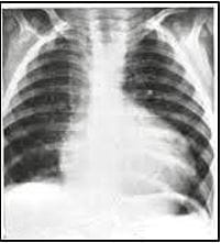
Figure 1: X-ray chest PA: cardiomegaly, reduced pulmonary blood flow.
pedia_724_02.JPGFigure 2: Twelve lead ECG: Left axis deviation, LVH.


On comprehensive sequential segmental analysis by color doppler echocardiography, there was situs solitus, levocardia, normal venoatrial connection (SVC, IVC and coronary sinus are opening into RA and 3 pulmonary veins are draining into LA (no anomalous pulmonary venous drainage was detected), atrioventricular concordance, with LA connecting to LV via bileaflet MV. RA and RV concordance was interrupted by the presence of prominent and conspicuous Tricuspid Atresia (Figure 3).
pedia_724_03.JPG
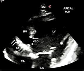
Figure 3: Apical 4CH view-Tricuspid atresia, large subaortic VSD, dilated LV, normal-sized RV. Horizontal arrows denote the presence of VSD, Oblique arrows denote Tricuspid atresia (TA).

Most strikingly, there was presence of DOLV with both great arteries arising exclusively from LV in a D-malposition arrangement, with aorta placed anterior and to the right of pulmonary artery, and pulmonary artery being posterior and to the left (Figure 4,5).

pedia_724_04.JPG
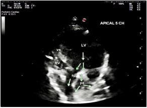
Figure 4: Apical 5CH view Double outlet left ventricle, D-malposition of great arteries, dilated LV ao, denotes Aorta pa, denotes Pulmonary Arteries.

pedia_724_05.JPG
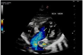
Figure 5: Apical 5CH view Double outlet left ventricle, Aorta is anterior and Pulmonary Artery is posterior, dilated LV, normal sized RV, hypoplasia of main and branch pulmonary arteries.

Mosiac pattern in pulmonary arteries is suggestive of turbulence in blood flow across mild pulmonary valvular stenosis.

A subaortic conus was distinctly detected (Figure 6). Meanwhile, pulmonary arteries were confluent and there was the presence of a left aortic arch. Mitral and aortic valves were normal, however, the pulmonary valve was domed and there was a presence of mild pulmonary stenosis with hypoplasia of pulmonary valve annulus, main and branch pulmonary arteries (Aortic valve annulus (D) 9.9 mm, PV annulus (D) 3.6 mm, main pulmonary artery (D) 3.0 mm, left pulmonary artery (D) 2.5 mm, right pulmonary artery (D) 2.6 mm). On CW doppler, peak pulmonary velocity was 1.95 m/sec with a peak gradient of 15.2 mmHg (Figure7). No infundibular obstruction was identified.
pedia_724_06.JPG
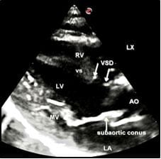
Figure 6: LAX view Subaortic VSD (moderate), characteristic subaortic conus, dilated LV.


pedia_724_07.JPG
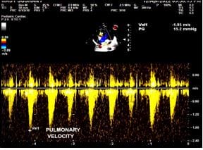
Figure 7: CW Doppler of the pulmonary artery:

There is mild pulmonary valvular stenosis-peak velocity 1.95 m/sec and peak gradient 15.2 mm hg. pedia_724_08.JPG
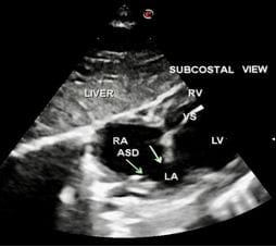
Figure 8: Subcostal View Large ostium secundum ASD denoted by oblique arrows.
pedia_724_09.JPG
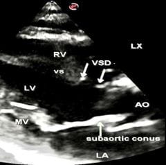
Figure 9: LAX view Subaortic VSD (moderate size) denoted by arrows.

Furthermore, there was a notable presence of Large ASD (Figure 8) and a moderate-sized VSD (Figure 9).

ASD was ostium secundum type of size 7.3 mm, with the right to left shunt. VSD was a subaortic membranous defect of size 4.3 mm, with a left to right shunt (Figure 10).

pedia_724_10.JPG
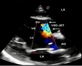
Figure 10: LAX view VSD jet is visualised with a left to right shunt across moderate size VSD.

VSD jet is denoted by arrows.

The peak velocity of the VSD jet was 1.34m/sec and the peak gradient was 7 mmHg. The left ventricle was dilated and the right ventricle was normal. No right ventricular hypoplasia was evident. The biventricular systolic functions were normal with an LVEF of 61%.

Discussion

Van Praagh et al [3]. extensively examined 109 cases of DOLV, based on autopsy material, personal communications and literature review. They concluded that DOLV is a unique anomaly in which the aorta and pulmonary arteries arise completely or predominantly from the morphologic left ventricle. According to this definition, the double-outlet left ventricle is a very heterogeneous entity, presenting wide variations in morphology [3]. The classification of DOLV is dependent on three principal criteria: a) the situs, based on a segmental approach; b) concordance or discordance of the atria, the ventricles and the great arteries; and c) associated cardiovascular and non-cardiovascular malformation. Due to the heterogeneity of morphological findings, Bharati et al


[7] remarked that it is not a precise entity. The explicit description of DOLV for treatment guidance was highlighted by Otero Cole et al [8].

In a noteworthy review of 126 cases by Gouton et al [9], it was reported that there was a greater incidence of levocardia (96%), atrial situs solitus (92.8%), subaortic interventricular communication (70%), and D-transposition (65%). Atrioventricular valve anomalies were observed in 30% of cases, mostly in the tricuspid valve and right ventricular hypoplasia is commonly associated. Van Praagh et al [3]. described DOLV with subaortic VSD to be the most common type of DOLV. Their series emphasized that the right anterior aorta was more frequent than the left. Correspondingly, Donald and William [10]. reported identical findings, describing that 83% of their cases of DOLV were associated with subaortic VSD and right anterior aorta with pulmonary stenosis. Likewise, our patient also had similar lesions in conformity with these authors [3, 9, 10]. Another author [11]. with a case report of classical DOLV, in a 4-year-old male child, diagnosed by cross-sectional Doppler echocardiography, and confirmed by cine angiocardiography, contended, that improvement in echocardiography techniques, allowed early diagnosis of DOLV, leading to an expeditious and early surgical intervention.

In our case, there was the presence of additional conspicuous tricuspid atresia, thus making it the rarest of a rare entity. On reviewing the literature only a few case reports of DOLV with tricuspid atresia could be found [12-15]. and severe hypoplasia of the right ventricle was present in the majority of these patients. However, in our case, there was the absence of right ventricular hypoplasia which is in agreement with Alehan et al [13], and according to them, the normal right ventricular development may result from a large VSD. Our patient had a moderate-sized VSD with normal right ventricular dimensions, which conforms with Alehan et al [13].

The presence or absence of conus, associated with DOLV has been cited by a few authors [16,17]. Multiple forms of conal presentations have been shown to occur in DOLV: subpulmonic, subaortic, bilateral present, and bilaterally absent [17]. Two case reports (a total of three patients) have identified subpulmonic or bilateral conus, in patients of DOLV undergoing biventricular repair [18, 19]. Manner et al reported subpulmonic, subaortic

and bilaterally absent conus associated with DOLV in their series of “Malpositions of the heart” [2]. It is obvious from these case reports that conus location does not follow a fixed pattern. Our case had a subaortic conus and subpulmonic conus was absent.

Another important case report [16]. revealed the absence of subpulmonic conus in two of their operated cases of DOLV by pulmonary root translocation for biventricular repair. The absence of subpulmonic conus causes a technically different procedure to be undertaken because it requires careful dissection of the deeply set pulmonary valve, from atrioventricular valve(s), with which it is in continuity. Otherwise, a traditional aortic valve replacement with pulmonary autograft could have been undertaken. This case report highlights the importance of conus location, and its bearing on the planning of surgical procedures, to be embarked upon.

Colour doppler echocardiography has substantially expanded the horizons for accurate non-invasive diagnosis of congenital cardiac defects including complex congenital heart disease [20]. Echocardiography not only provides additional information but also improves the overall specificity of two-dimensional (2-D) echocardiography.

Traditionally, DOLV has been difficult to diagnose precisely. Previously angiography was considered indispensable and the diagnosis was often concluded by the surgeon or pathologist [21]. Hence, DOLV may have been underdiagnosed. Today, cardiac MRI as an adjunct to echocardiography provides reliable and sophisticated diagnostic techniques. However, our country, belongs to a low-income group of nations and cardiac MRI facility is extremely limited moreover, adequate trained academic teaching faculty of cardiac MRI, particularly with the expertise of reading and reviewing paediatric cardiology cases, are lacking. Moreover, it is time-consuming and expensive, which are other important hindrances to the use of this advanced technology in our motherland. Our institution does not possess a cardiac MRI. The innovative technology of 4Dimensional XStrain Echocardiography utilizes LV border tracking, by fusing speckle tracking information obtained from standard apical 2CH, 3CH and 4CH views, and delivering a more complete picture of cardiac function. This advanced technology merges


Tomtec GMBH’s 3D/4D rendering and BeutelTM computation capabilities [22]. This tool, relying on the high spatial and temporal resolution of 2D imaging, addresses and resolves the major limitations currently related to the use of full-volume 3D STE [23,24]. We employed 4Dimensional XStrain Echocardiography, in a detailed sequential chamber analysis of the current case report.

Strengths and Limitations: To attain a clinching diagnosis, our strength was a comprehensive color doppler echocardiography, performed by the author himself, employing the 4Dimensional XStrain echocardiography system of Esaote, Italy. Because of the excellent resolution of images, we were able to negotiate the complexities of lesions and come to a definitive diagnosis. The limitations were the absence of cardiac MRI to support our diagnosis, because of the reasons already explained above.

Conclusion

We have presented an exceedingly rare case report of DOLV, Tricuspid Atresia, Large Ostium Secundum ASD, Moderate size subaortic VSD, D-malposition of great arteries, and mild pulmonary valvular stenosis, detected and evaluated by sequential chamber approach, employing transthoracic color doppler echocardiography.

Acknowledgement: We acknowledge the incredible efforts of Ms Shubham Kacker to edit the manuscript, despite her hectic work schedule and intense family responsibilities.

Reference

01. Bengur AR, Snider AR, Peters J, Merida-Asmus L. Two-dimensional echocardiographic features of double outlet left ventricle. J Am Soc Echocardiogr. 1990 Jul-Aug; 3(4):320-5. [Crossref][PubMed][Google Scholar]

02. Manner J, Seidl W, Steding G. Embryological observations on the formal pathogenesis of double-outlet left ventricle with a right-ventricular infundibulum. Thorac Cardiovasc Surg. 1997 Aug; 45(4):172-7. [Crossref][PubMed][Google Scholar]

03. Adams, Forrest H. , and George C. Emmanouilides. Moss’ heart disease in infants, children and adolescents. Moss’ Heart disease in infants, children and adolescents. 1983.795-795 [Crossref][PubMed][Google Scholar]

04. Menon, Shaji C. , and Donald J. Hagler. Double-outlet left ventricle: diagnosis and management. Current treatment options in cardiovascular medicine 2008; 10(5):448-452 [Crossref][PubMed][Google Scholar]

05. Sakakibara, S. Both great vessels arising from left ventricle. “Bull Heart Inst Jpn” 1967; 11:66-86. . [Crossref][PubMed][Google Scholar]

06. Lilje C, Weiss F, Lacour-Gayet F, Razek V, Ntalakoura K, Weil J, et al. Images in Cardio-vascular medicine. Double-outlet Left Ventricle. Circulation 2007 Jan 23;, 115(3):e36-7. [Crossref][PubMed][Google Scholar]

07. Bharati S, Lev M, Stewart R, McAllister HA, Jr, Kirklin JW. The morphologic spectrum of double outlet left ventricle and its surgical significance. Circulation 1978 Sep; 58: 558-65. . [Crossref][PubMed][Google Scholar]

08. Coto EO, Jimenez MQ, Castaneda AR, Rufilanchas JJ, Deverall PB. Double outlet from chamber of left ventricular morphology. Br Heart J. 1979 Jul; 42(1):15-21. [Crossref][PubMed][Google Scholar]

09. Gouton M. , et al. Double outlet left ventricle: a rare and unusual cardiopathy. Apropos of 7 new cases. Archives des Maladies du Coeur et des Vaisseaux 1966; 89(5): 553-559 [Crossref][PubMed][Google Scholar]

10. Van Praagh, R. Double-outlet left ventricle. Heart Diseases in infants, children and Adolescents (1989); 461-480. . [Crossref][PubMed][Google Scholar]

11. Mohan JC, Agarwala R, Arora R. Double outlet left ventricle with intact ventricular septum; a cross-sectional and Doppler echocardiographic diagnosis. Int J Cardiol. 1991 Dec; 33(3); 447-9. [Crossref][PubMed][Google Scholar]

12. Rocha, Isaura Elaine Goncalves, Isaura Corina Pazin and Lilian Maria Lopes. Tricuspid atresia and double outlet left ventricle: A Rare association in adulthood. Arq Bras Cardiol: Imagem cardiovasc. 2017; 30(1):31-35. [Crossref][PubMed][Google Scholar]

13. Alehan D, Dogan OF, Ozkutlu S. Echocardiographic diagnosis of an extremetly rare case with double-outlet left ventricle, tricuspid atresia and two balanced ventricles: Pediatr Cardiol


2007 Sep-Oct;28(5): 418-9. . . [Crossref][PubMed][Google Scholar] [Crossref][PubMed][Google Scholar]

14. Vaseenon T, Diehl AM, Mattioli L. Tricuspid atresia with double-outlet left ventricle and bilateral conus. Chest. 1978 Dec; 74(6): 676-9. [Crossref][PubMed][Google Scholar]

15. Lopes LM, Rangel PI, Soraggi AM, Furlanetto BH, Furlanetto G: Double outlet left ventricle. Echocardiographic diagnosis: Arq Bras Cardiol. 2001 Jun; 76(6):511-6. . [Crossref][PubMed][Google Scholar]

16. McElhinney DB, Reddy VM, Hanley FL. Pulmonary root translocation for biventricular repair of double-outlet left ventricle with absent subpulmonic conus. J Thorac Cardiovasc Surg. 1997 Sep; 114(3):501-3. [Crossref][PubMed][Google Scholar]

17. Hagler, D. J. Double-outlet right ventricle and double outlet left ventricle. “Moses and Adams’ Heart Disease in infants, Children, and Adolescents (2008). pp.: 1100-1127 [Crossref][PubMed][Google Scholar]

18. DeLeon SY, Ow EP, Chiemmongkoltip P, Vitullo DA, Quinones JA, Fisher EA, et al. Alternatives in biventricular repair of double-outlet left ventricle. Ann Thorac Surg 1995 Jul; 60(1):213-6. . [Crossref][PubMed][Google Scholar]

19. Chiavarelli M, Boucek MM, Bailey LL. Arterial correction of double outlet left ventricule by pulmonary artery translocation. Ann Thorac Surg. 1992 Jun; 53(6):1098-100. [Crossref][PubMed][Google Scholar]

20. Huhta JC, Hagler DJ, Seward JB, Tajik AJ, Julsrud PR, Ritter DG. Two-dimensional echocardiographic assessment of dextrocardia: a segmental approach. AM J Cardiol. 1982 Dec; 50(6):1351-60. [Crossref][PubMed][Google Scholar]

21. Galal O, Hatle L, AL Halees Z. Changes of management in a patient with double outlet left ventricle. Cardiol young 1999 Nov; 9(6):602-5. . [Crossref][PubMed][Google Scholar]

22. Muraru D, Niero A, Rodriguez-Zanella H, Cherata D, Badano L. Three-dimensional speckle-tracking echocardiography: benefits and limitations of integrating myocardial

mechanics with three-dimensional imaging. Cardiovasc Diagn Ther. 2018 Feb; 8(1):101-117. [Crossref][PubMed][Google Scholar]

23. Carreras F, Garcia-Barnes J, Gil D, Pujadas S, Li CH, Suarez-Arias R, et al. Left ventricular torsion and longitudinal shortening: two fundamental components of myocardial mechanics assessed by tagged cine-MRI in normal subjects. Int J Cardiovasc Imaging. 2012 Feb; 28(2):273-84. [Crossref][PubMed][Google Scholar]

24. Takahashi K, Al Naami G, Thompson R, Inage A, Mackie AS, Smallhorn JF. Normal rotational torsion and untwisting data in children, adolescents and young adults. J Am Soc Echocardiogr. 2010 Mar; 23(3):286-93. [Crossref][PubMed][Google Scholar]