E-ISSN:2349-3267
P-ISSN:2349-5499

Case Report

DCRV

Pediatric Review - International Journal of Pediatric Research

2022 Volume 9 Number 4 July-August
Publisherwww.medresearch.in

Double-Chambered Right Ventricle with Large Apical VSD- a Rare Case Report

Mehrotra A.1*, Sharma A.2, Singh S.3, Kacker S.4
DOI: https://doi.org/10.17511/ijpr.2022.i04.03

1* Akhil Mehrotra, Chief, Non-Invasive Cardiologist, Pediatric and Adult Cardiology, Prakash Heart Station, D-16, Nirala Nagar, Lucknow , U.P., India.

2 Ajay Sharma, Senior Echocardiography Technician, Pediatric and Adult Cardiology, Prakash Heart Station, D-16, Nirala Nagar, Lucknow , U.P., India.

3 Shwati Singh, Senior Echocardiography Technician, Pediatric and Adult Cardiology, Prakash Heart Station, D-16, Nirala Nagar, Lucknow , U.P., India.

4 Shubham Kacker, Senior Coordinator, Project Management Office, Tech Mahindra, Noida, U.P., India.

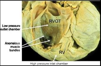
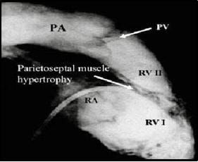
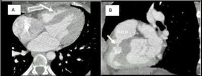
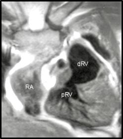
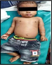
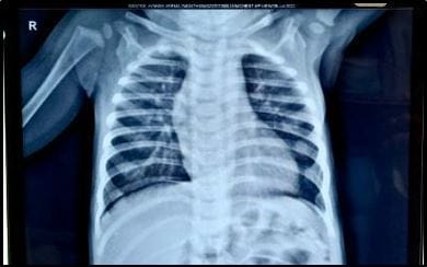
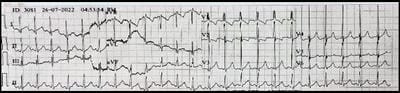
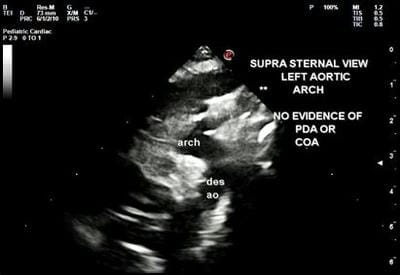
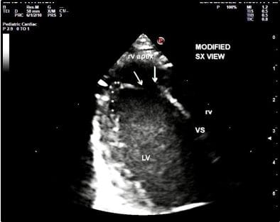
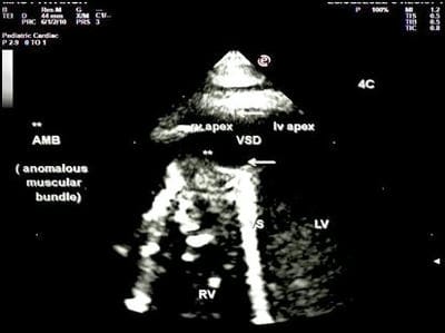
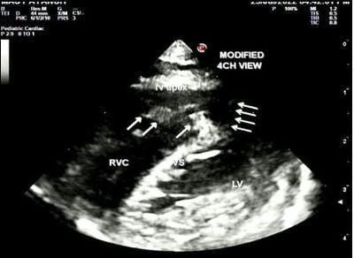
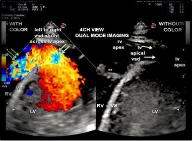
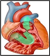
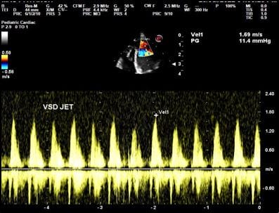
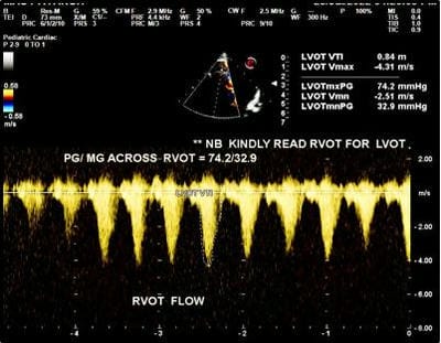
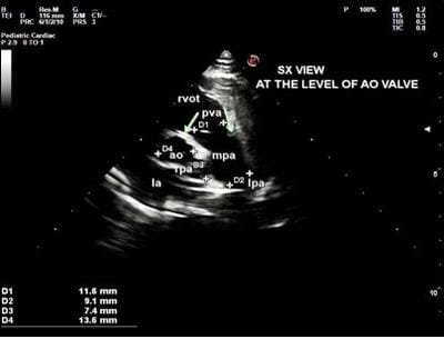
Double-chambered right ventricle (DCRV) is a rare condition seen in only 0.5 - 2.0% of all cases of congenital heart disease (CHD). An isolated DCRV is very rare, while approximately 80-90% of DCRV cases are associated with various other congenital heart defects, with VSD, in particular, a perimembranous type VSD, being the most common. In DCRV right ventricle is separated into a proximal high-pressure and distal low-pressure chamber. It can be caused either by the presence of an anomalous muscle bundle (AMB), by hypertrophy of endogenous trabecular tissue, or occasionally by an aberrant moderator band. DCRV is characterised by intraventricular pressure gradients greater than 20 mmHg, turbulent flow patterns in the ventricle, and increased pulmonary blood flow. Currently, the methods for detection of DCRV with VSD are Colour echocardiography, Cardiac catheterization, Cardiac CT and Cardiac MRI. This anomaly is often diagnosed during childhood and adolescence, while very few are found in adults. Here, we are presenting an extremely rare case report of a 7-month-old male child afflicted with symptomatic DCRV, unusually associated with a large apical muscular VSD.

Keywords: Double-chambered right ventricle, Large Apical VSD, 4Diemensional XStrain Echocardiography, Anomalous Muscle bundle

Corresponding Author How to Cite this Article To Browse
Akhil Mehrotra, Chief, Non-Invasive Cardiologist, Pediatric and Adult Cardiology, Prakash Heart Station, D-16, Nirala Nagar, Lucknow , U.P., India.
Email:
Akhil Mehrotra, Ajay Sharma, Shwati Singh, Shubham Kacker, Double-Chambered Right Ventricle with Large Apical VSD- a Rare Case Report. Pediatric Rev Int J Pediatr Res. 2022;9(4):25-33.
Available From
https://pediatrics.medresearch.in/index.php/ijpr/article/view/726

Manuscript Received Review Round 1 Review Round 2 Review Round 3 Accepted
2022-08-02 2022-08-04 2022-08-11 2022-08-18 2022-08-25
Conflict of Interest Funding Ethical Approval Plagiarism X-checker Note
Nil Nil Yes 18%

© 2022by Akhil Mehrotra, Ajay Sharma, Shwati Singh, Shubham Kackerand Published by Siddharth Health Research and Social Welfare Society. This is an Open Access article licensed under a Creative Commons Attribution 4.0 International License https://creativecommons.org/licenses/by/4.0/ unported [CC BY 4.0].

Introduction

A double-chambered right ventricle (DCRV) is a heart defect, in which the right ventricle (RV) is separated into a proximal high-pressure (anatomically lower) chamber and distal low-pressure (anatomically higher) chamber [1,2] (Figure 1) it can be caused either by the presence of anomalous muscle bands, by hypertrophy of endogenous trabecular tissue, or occasionally by an aberrant moderator band.

However, DCRV has also been reported to develop postnatally due to progressive hypertrophic changes in the crista supraventricularis or other muscular structures within the RV [3, 4]

pedia_726_01.JPG

Figure 1: The photograph shows a double-chambered right ventricle (RV) with anomalous muscle bundles located below the infundibulum, which divides the RV into a high-pressure inlet chamber and a low-pressure outlet chamber. (RVOT = right ventricular outflow tract.).

DCRV was first described by Peacock in 1867 as a constriction of the proximal portion of the infundibulum [5].

In 1909, Keith described a muscular shelf extending into the apex of the ventricle. Brock later described, in 1957, an infundibular muscular obstruction in the setting of tetralogy of Fallot [6, 7]. Outflow obstruction was observed to be directly caused by anomalous muscle tissue by Tsifutis in 1961 and was first surgically corrected by Lucas et al. in 1962 through a partial ventriculotomy [8, 9].

DCRV is characterized by intraventricular pressure gradients greater than 20 mmHg, turbulent flow patterns in the ventricle, and increased pulmonary flow [2].

Currently, the methods for detection of DCRV with VSD, besides echocardiography are Cardiac catheterization, cardiac CT and cardiac MRI (Figure 2, 3, 4).

DCRV is a rare condition seen in only 0.5-2.0% of all cases of congenital heart disease and is most frequently encountered in infants and children [2, 3, 10]. While cases have been found in adults, these might be due to missed diagnoses during infancy rather than novel onset later in life [2, 11].

An isolated DCRV is very rare, representing only in 6.2% of the patients, while approximately 80-90% of the DCRV cases are associated with various congenital heart defects, with ventricular septal defect (VSD), in particular, a perimembranous type VSD, being the most common. According to Hoffman, the most frequent associated congenital heart defect in DCRV patients was VSD, which accounted for 84.4%, followed by membranous subaortic stenosis (31.3%) [2].

DCRV can also be an associated anomaly of Williams’s syndrome. [12].This anomaly is often diagnosed during childhood and adolescence, while very few were found in adults [13]. Here, we are presenting a rare case report of a 7-month male child suffering from DCRV along with the co-existence of unorthodox and non-conventional large apical muscular VSD.

pedia_726_02.JPG

Figure 2: Cardiac catheterisation and RV angiography in a patient with DCRV, in lateral view the two chambers within the right ventricle, divided by hypertrophied septoparietal musculature. RV |,proximal RV chamber,RV ||, distal RV chamber , RA - right atrium, PA - pulmonary artery, PV - pulmonary valve.


pedia_726_03.JPG

Figure 3: Cardiac CT in DCRV. Panels A and B demonstrate axial and sagittal views respectively, on CTA.

The arrows demarcate an AMB crossing from the interventricular septum to the RV free wall, consistent with DCRV.

pedia_726_04.JPG

Figure 4: Turbo- spin-echo MR image demonstrates a muscle band subdividing the RV into a proximal (lower) and distal (upper) chamber.

RA - right atrium, pRV - proximal RV chamber and dRV - distal RV chamber.

Case Report

A 7 Month male child was referred to us for evaluation of a heart murmur from a private pediatric hospital.

The parents were extremely cooperative while enumerating the details of the history. The child was a full-term normal delivery from a multipara woman of 23 years of age, delivered at a private hospital with normal birth weight, and was normal at birth.

The vaccination of mother and child was appropriately carried out, at the date of presentation to us.

There was no history of maternal risk factors for CHD (morbid obesity, diabetes, febrile illness, smoking, alcohol intake, teratogenic drugs use, or radiation exposure).

On deep interrogation, the parents informed that the child was having recurrent chest infections, failure to thrive and chest retractions during chest infections.

On clinical examination he was weighing 5.5 kg, weight 42 cm, BP 90/60 mmHg, Pulse rate 113/min, RR 25/min and SPO2 99% at room air. The child was of average built mildly tachypneic and irritable (Figure 5), without any evident chest retractions, cyanosis, clubbing or signs of heart failure. There was an absence of musculoskeletal anomalies. All the peripheral pulses were normally palpable without any radio-femoral delay.

Meanwhile, the respiratory system, central nervous system and abdomen were also examined and no abnormality was detected.

pedia_726_05.JPG

Figure 5: In our case - a 7-month male child afflicted with DCRV and Large apical VSD.


pedia_726_06.JPG

Figure 6: X-ray Chest PA view - There is cardiomegaly with increased pulmonary blood flow

On cardiovascular examination, there was the presence of a harsh grade 3/6 ejection systolic murmur, heard best at the right sternal edge and Left second intercostal space, adjacent to the sternum. No ejection click was audible. IInd heart sound was normal. Chest X-ray PA view (Figure 6),

Showed cardiomegaly with signs of increased pulmonary blood flow. Resting 12 lead ECG (Figure 7) revealed sinus tachycardia (ventricular rate~ 115/min), with partial Right bundle branch block (RBBB) and a normal QRS axis. Partial RBBB may suggest presence of right ventricular hypertrophy or increased right ventricular systolic pressure (RVSP).

Figure 7: Resting 12 lead ECG - There is partial RBBB, Sinus Tachycardia (VR -115/min) and normal QRS axis.

Comprehensive 4Diemensional XStrain Echocardiography

The patient underwent colour echocardiography by 4Dimensional XStrain Echocardiography system in supine and left lateral decubitus posture, and detailed sequential chamber analysis was done from subcostal, parasternal long axis, parasternal short axis, apical four-chamber and suprasternal views.

There was levocardia, situs solitus, atrioventricular concordance, ventricular-arterial concordance, concordant D-bulboventricular loop, normally related great arteries, confluent pulmonary arteries and left aortic arch (Figure 8). There was the absence of patent ductus arteriosus or coarctation of the aorta.

pedia_726_08.JPG

Figure 8: Suprasternal view - There are left aortic arch without any evidence of COA or PDA.

In the modified SX view (Figure 9), a distinctive large apical muscular VSD of size 7.4 mm was precisely delineated.

Moreover, in the apical 4CH and modified 4CH view, we could clearly demarcate an apical VSD communicating with the RV apex, along with a notable and pronounced anomalous muscle bundle (AMB) (Figures 10 and 11 respectively), lying just proximal to the apical VSD.

This AMB divided RV into two chambers: 1) small RV apical chamber (lower chamber) and 2) normal-sized basal RV chamber (upper chamber), consisting of RV inflow and RV inflow and RV cavity.

RV apex was resembling, as if it is a continuation of the apical region of LV, because of the presence of large conspicuous VSD. 

pedia_726_09.JPG

Figure 9: Modified SX view - Arrows demarcate an apical large VSD communicating with RV apex, ** asterix - denotes LV apex.


pedia_726_10.JPG

Figure 10: Apical 4CH view- Arrows indicate large apical VSD causing free communication between LV and RV apex. ** Asterisk exemplifies the presence of peculiar AMB.

pedia_726_11.JPG

Figure 11: Modified 4CH view - 4 horizontal arrows illustrate large apical VSD, 3 vertical arrows denote AMB.

pedia_726_12.JPG

Figure 12: 4CH view- with dual mode imaging. In the left black and white panel Large apical VSD is indicated by arrows and AMB is denoted by an asterisk **.

Color Doppler echocardiography (CDE) on dual mode imaging revealed spectacular and magnificent echo images with laminar flow across large apical VSD and turbulent, mosaic pattern flow across AMB (Figure 12)

In the right coloured panel, 2 angulated arrows point to the non-turbulent left to right flow across VSD and 3 angulated arrows indicate a highly turbulent mosaic pattern flow across AMB.

On CDE the direction of blood flow was from left to right across the VSD and subsequently from the lower, apical RV chamber to the upper, basal RV chamber across a severely restricting AMB.

This direction of flow is in contrast to the usual flow in DCRV- from the RV base to the RV apex (Figure 13).

pedia_726_13.JPG

Figure 13: An anatomical illustration of DCRV showing a pattern of blood flow (in green colour) in a usual patient.

The image distinctly shows that the RV blood flows from a proximal high-pressure chamber (basal chamber) into a distal low-pressure chamber (apical chamber) via a restrictive AMB

On CW doppler, the peak velocity of the VSD jet was 1.69 m/sec with a peak gradient of 11.4 mmHg (Figure 14)


pedia_726_14.JPG

Figure 14: CW Doppler flow across large apical VSD, in our patient.

There is a peak gradient of 11.4 mm hg with a peak velocity of 1.69 m/sec.

However, on CW doppler evaluation across AMB, a peak velocity of 4.3 m/sec with a peak/mean gradient of 74.2/32.9 mmHg was discerned (Figure 15).

pedia_726_15.JPG

Figure 15: CE Doppler flow velocity across AMB (from RV apex to RV cavity).

The peak/ mean gradient across AMB was 74.2/32.9 mmhg.

Furthermore, in the SX view at the level of the aortic valve, the pulmonary valve was normal and the main and branch pulmonary arteries were dilated (Figure 16).

pedia_726_16.JPG

Figure 16. SX View at the level of AO valve.

The pulmonary valve is normal and the main and branch pulmonary arteries are dilated. RVOT - right ventricular outflow tract, PVA - pulmonary valve annulus, MPA - main pulmonary artery, LPA - left pulmonary artery, RPA - right pulmonary artery, AO - aorta.

There was concentric hypertrophy of the RV without any dilatation and normal RV systolic function. The left ventricle was of normal size and the systolic function was also normal, LVEF 69%.

Because of the presence of a large apical VSD with restrictive AMB, with significant symptoms, the parents were advised that the child should undergo corrective surgery, in the form of patch closure of VSD and resection of AMB, at a tertiary care centre.

Discussion

DCRV accounts for 0.5 -2% of congenital heart disease and occurs in as many as 10% of patients with VSD [2]. The male-to-female ratio is 2:1. No inheritance pattern or risk factors are described. Sporadic cases have been reported in patients with Down and Noonan Syndromes. Associated congenital cardiac abnormalities are found in 80-90% of cases. Isolated DCRV is exceptionally rare [14]. VSD is the most common defect, the next being pulmonary stenosis. Other associations are double outlet right ventricle, tetralogy of Fallot, anomalous pulmonary venous drainage, transposition of the great arteries, pulmonary atresia with an intact ventricular septum, and Ebstein anomaly [15]. VSD is usually large, perimembranous and opens into the high-pressure proximal chamber but it may open into the distal


chamber also. As the obstruction of DCRV worsens, associated VSD may progressively become smaller. Few authors have reported that asymptomatic adults with AMB and intact ventricular septum may have had VSD that underwent spontaneous closure [16].

Research on spatial relations between VSD and anomalous muscle band revealed that the VSD was proximal to the obstructing muscle bundle in 62% and distal to the bundle in 38% of patients [17]. However, in the literature, the relation between VSD and muscle band was not indicated in most instances. In some series, the VSD was noted to open to the proximal chamber in all cases: while in others, it opened into the distal chamber thus acting as an extension of the left ventricle [17]. In general, VSD was proximal to the anomalous muscle band in 2/3 cases [17].

For diagnosis transthoracic echocardiography (TTE) may be insufficient, so transesophageal echocardiography (TTE) is strongly advised for both children and adults, particularly in the presence of right ventricular hypertrophy on electrocardiogram. It can be difficult to obtain an image owing to the proximity of the right ventricular outflow tract to the transducer. Colour flow Doppler identifies the site of obstruction by the appearance of a mosaic pattern where the high-velocity flow originates. [5, 8] Cardiac catheterisation may be performed to confirm the diagnosis. Pressure in the distal chamber is equal to pulmonary artery unless there is associated pulmonary valve stenosis. Right ventricular angiography showing filling defects within the right ventricle, between the outflow and inflow areas, confirms the diagnosis. Left ventriculography is performed for associated VSD or subaortic stenosis. Cardiac magnetic resonance can visualize RV anatomy, obstructing muscular bundles together with a determination of the pressure gradient [18].

AMB when found in the right ventricular apical region are generally of little functional significance. On the other hand, a muscle bundle situated across the main channel of the right ventricular cavity can cause haemodynamic disturbances, especially when it becomes hypertrophied. Usually, the anomalous muscle bands divide the right ventricle into 2 chambers with a proximal high-pressure chamber connected to the inflow and a distal low-pressure chamber connected to the RV outflow [2]. In our

case, the muscle band in the RV apex divided the right ventricle into 2 chambers: 1) proximal chamber consisting of both RV inflow and RV cavity and 2) distal small RV apical chamber which was in communication with the left ventricular cavity via large non-restrictive apical muscular ventricular septal defect. The haemodynamic consequences and symptoms of the large non-restrictive ventricular septal defect were curtailed by the restrictive muscle band in the right ventricle. Overall clinically, echocardiographically and hemodynamically condition behaved like restrictive ventricular septal defect with left to right shunt.

The small apical muscular ventricular septal defect can close spontaneously but larger defects often persist and need treatment [19]. Though a number of cases of anomalous muscle band with typical DCRV have been reported in the literature; our case is atypical because of the presence of a proximal low-pressure chamber and a distal high-pressure RV apical chamber, which is just in contrast to the usual cases reported in the literature [5].

There was been one similar case reported in the literature, where an 11-year-old asymptomatic girl was found to have an apical muscular ventricular septal defect that was large but behaved like a small defect because of the restrictive flow across the anomalous muscle bundles in the right ventricular apex, and the patient was kept under medical follow up [20].

In the literature search, we also found another anatomically similar case wherein a 48-year-old male, echocardiography and magnetic resonance imaging revealed a large apical ventricular septal defect with separation of the right ventricular apex from the remaining RV by excessive trabeculations, thereby leading to the elimination of left to right shunt across the VSD. Physiologically there was no hemodynamic disturbance so the patient was kept under medical follow-up [21].

Therefore, this type of apical VSD along with apical muscle bands constitutes a rare and distinct type of morphology and physiology, and treatment needs to be individualized based on the hemodynamic disturbance it causes. Our patient was significantly symptomatic, hence we advised the parents of the child to seek an opinion from the department of Pediatric Cardiac Surgical unit of a tertiary care centre, for surgical resection of AMB with patch closure of the apical VSD.


Conclusion

Though several cases of DCRV with AMB have been reported in the literature, our case is unique in the sense that AMB was dividing the RV into two chambers: 1) a small RV apical chamber and 2) normal sized, basal RV chamber consisting of RV inflow and RV cavity.

Importantly, there was laminar, non-turbulent left to right flow across VSD with subsequent flow across AMB, from distal apical RV chamber to proximal basal RV chamber. This is in contrast to other reports published in the literature.

Acknowledgement: We are immensely grateful to Ms Shubham Kacker for her valuable and constructive suggestions, throughout the process of editing the manuscript. Her willingness to give her precious time so generously is deeply appreciated. We would also like to express our gratitude to Mrs Laxmi Kannoujia for her untiring efforts to repeatedly type the article with noteworthy zeal and enthusiasm.

Reference

01. Nagashima M, Tomino T, Satoh H, Nakata T, Ohtani T, Saito H. Double-chambered right ventricle in adulthood. Asian Cardiovasc Thorac Ann. 2005 Jun;13(2):127-30. doi: 10.1177/021849230501300206 [Crossref][PubMed][Google Scholar]

02. Hoffman P, Wójcik AW, Rózański J, Siudalska H, Jakubowska E, Włodarska EK, et al. The role of echocardiography in diagnosing double chambered right ventricle in adults. Heart. 2004 Jul;90(7):789-93. doi: 10.1136/hrt.2003.017137 [Crossref][PubMed][Google Scholar]

03. Restivo A, Cameron AH, Anderson RH, Allwork SP. Divided right ventricle: a review of its anatomical varieties. Pediatr Cardiol. 1984 Jul-Sep;5(3):197-204. doi: 10.1007/BF02427045 [Crossref][PubMed][Google Scholar]

04. Wang JK, Wu MH, Chang CI, Chiu IS, Chu SH, Hung CR, et al. Malalignment-type ventricular septal defect in double-chambered right ventricle. Am J Cardiol. 1996 Apr 15;77(10):839-42. doi: 10.1016/S0002-9149(97)89178-3 [Crossref][PubMed][Google Scholar]

05. Alva C, Ho SY, Lincoln CR, Rigby ML, Wright A, Anderson RH. The nature of the obstructive muscular bundles in double-chambered right ventricle. J Thorac Cardiovasc Surg. 1999 Jun;117(6):1180-9. doi: 10.1016/s0022-5223(99)70258-8 [Crossref][PubMed][Google Scholar]

06. Brock RC. The anatomy of congenital pulmonary stenosis. London: Cassel; 1957. 42-51. [Crossref][PubMed][Google Scholar]

07. Keith, Arthur. The Hunterian lectures on malformations of the heart. 1. " Lancet 2 (1909): 519-523. [Crossref][PubMed][Google Scholar]

08. LUCAS RV Jr, VARCO RL, LILLEHEI CW, ADAMS P Jr, ANDERSON RC, EDWARDS JE. Anomalous muscle bundle of the right ventricle. Hemodynamic consequences and surgical considerations. Circulation. 1962 Mar;25:443-55. doi: 10.1161/01.cir.25.3.443 [Crossref][PubMed][Google Scholar]

09. Tsifutis, A. A. , H. Arvidsson, and AF HARTMANN. "2 CHAMBERED RIGHT VENTRICLE-REPORT ON 7 PATIENTS." Circulation 24.4 (1961): 1058 [Crossref][PubMed][Google Scholar]

10. Folger GM Jr. Right ventricular outflow pouch associated with double-chambered right ventricle. Am Heart J. 1985 May;109(5 Pt 1):1044-9. doi: 10.1016/0002-8703(85)90248-0 [Crossref][PubMed][Google Scholar]

11. Park JG, Ryu HJ, Jung YS, Kim KJ, Lee BR, Jung BC, Kang H. Isolated double-chambered right ventricle in a young adult. Korean Circ J. 2011 May;41(5):272-5. doi: 10.4070/kcj.2011.41.5.272 [Crossref][PubMed][Google Scholar]

12. Yuan SM. Congenital heart defects in Williams syndrome. Turk J Pediatr. 2017;59(3):225-232. doi: 10.24953/turkjped.2017.03.001 [Crossref][PubMed][Google Scholar]

13. Oliver JM, Garrido A, González A, Benito F, Mateos M, Aroca A, et al. Rapid progression of midventricular obstruction in adults with double-chambered right ventricle. J Thorac Cardiovasc Surg. 2003 Sep;126(3):711-7. doi: 10.1016/s0022-5223(03)00044-8 [Crossref][PubMed][Google Scholar]

14. Park JI, Kim YH, Lee K, Park HK, Park CB. Isolated double-chambered right


ventricle presenting in adulthood. Int J Cardiol. 2007 Oct 18;121(3):e25-7. doi: 10.1016/j.ijcard.2006.11.024 [Crossref][PubMed][Google Scholar]

15. Lascano ME, Schaad MS, Moodie DS, Murphy D Jr. Difficulty in diagnosing double-chambered right ventricle in adults. Am J Cardiol. 2001 Oct 1;88(7):816-9. doi: 10.1016/s0002-9149(01)01862-8 [Crossref][PubMed][Google Scholar]

16. Matina D, van Doesburg NH, Fouron JC, Guérin R, Davignon A. Subxiphoid two-dimensional echocardiographic diagnosis of double-chambered right ventricle. Circulation. 1983 Apr;67(4):885-8. doi: 10.1161/01.cir.67.4.885 [Crossref][PubMed][Google Scholar]

17. Hubail ZJ, Ramaciotti C. Spatial relationship between the ventricular septal defect and the anomalous muscle bundle in a double-chambered right ventricle. Congenit Heart Dis. 2007 Nov-Dec;2(6):421-3. doi: 10.1111/j.1747-0803.2007.00135.x [Crossref][PubMed][Google Scholar]

18. Kilner PJ, Sievers B, Meyer GP, Ho SY. Double-chambered right ventricle or sub-infundibular stenosis assessed by cardiovascular magnetic resonance. J Cardiovasc Magn Reson. 2002;4(3):373-9. doi: 10.1081/jcmr-120013300 [Crossref][PubMed][Google Scholar]

19. Shirali GS, Smith EO, Geva T. Quantitation of echocardiographic predictors of outcome in infants with isolated ventricular septal defect. Am Heart J. 1995 Dec;130(6):1228-35. doi: 10.1016/0002-8703(95)90147-7 [Crossref][PubMed][Google Scholar]

20. Khositseth A. Large apical muscular ventricular septal defect: asymptomatic due to anomalous muscle bundles in the right ventricle. Congenit Heart Dis. 2007 Jan-Feb;2(1):70-3. doi: 10.1111/j.1747-0803.2007.00075.x [Crossref][PubMed][Google Scholar]

21. Grittner F, Daehnert I, Gutberlet M, Lurz P. Late diagnosis of a congenital apical ventricular septal defect with complete closure by right ventricular trabeculations. Eur Heart J Cardiovasc Imaging. 2014 Feb;15(2):230. doi: 10.1093/ehjci/jet170 [Crossref][PubMed][Google Scholar]