E-ISSN:2349-3267
P-ISSN:2349-5499

Case Report

Hydranencephaly

Pediatric Review - International Journal of Pediatric Research

2022 Volume 9 Number 5 September-October
Publisherwww.medresearch.in

Hydranencephaly – A rare case of a neglected child with enlarging head size

Patel S.1*, Sandeep B.2, Kiran B.3
DOI: https://doi.org/10.17511/ijpr.2022.i05.02

1* Shefali Patel, Assistant Professor, Department of Paediatrics, East Point College of Medical Sciences and Research Centre, Bangalore, Karnataka, India.

2 B Sandeep, Professor, Department of Paediatrics, East Point College of Medical Sciences and Research Centre, Bangalore, Karnataka, India.

3 B Kiran, HOD & Professor, Department of Paediatrics, East Point College of Medical Sciences and Research Centre, Bangalore, Karnataka, India.

Hydranencephaly is a rare congenital abnormality characterized by the absence and replacement of the cerebral hemisphere by cerebrospinal fluid. Here we report a rare case of a child who presented very late at 2 years of age with a massively enlarged head size with spasticity of all four limbs, suspected of hydrocephalous at first while on further work up diagnosed to have hydranencephaly. This report is aimed to make clinicians aware of a rare condition which can present very late even up to 2 years of age.

Keywords: Hydranencephaly, cerebrospinal fluid, hydrocephalous, spasticity

Corresponding Author How to Cite this Article To Browse
Shefali Patel, Assistant Professor, Department of Paediatrics, East Point College of Medical Sciences and Research Centre, Bangalore, Karnataka, India.
Email:
Shefali Patel, B Sandeep, B Kiran, Hydranencephaly – A rare case of a neglected child with enlarging head size. Pediatric Rev Int J Pediatr Res. 2022;9(5):34-37.
Available From
https://pediatrics.medresearch.in/index.php/ijpr/article/view/728

Manuscript Received Review Round 1 Review Round 2 Review Round 3 Accepted
2022-09-01 2022-09-02 2022-09-09 2022-09-16 2022-09-23
Conflict of Interest Funding Ethical Approval Plagiarism X-checker Note
Nil Nil Yes 16%

© 2022by Shefali Patel, B Sandeep, B Kiranand Published by Siddharth Health Research and Social Welfare Society. This is an Open Access article licensed under a Creative Commons Attribution 4.0 International License https://creativecommons.org/licenses/by/4.0/ unported [CC BY 4.0].

Introduction

Hydranencephaly is a rare encephalopathy. It is characterized by either the absence of cerebral hemispheres or represented by a membranous sac containing cerebrospinal fluid (CSF) with dispersed remnants of frontal, temporal or occipital centre over the membrane [1]. The thalamus, cerebellum and brainstem are relatively intact [2]. The cranial cavity may have remnants of glial tissue and ependyma, especially along the falx and close to the diencephalon [3]. The incidence is around 1 in 10,000 births [4].

Case report

A 2year old female child born of a 2nd degree consanguineous marriage, first in order of two with maternal age of 21 years and paternal age of 26years at the time of conception. Other  child is 4 months old and is normal. mother had not registered for an antenatal clinic. At around 32weeks of gestation, the mother had excessive pain abdomen and had gone to the tertiary centre where they had suspected severe oligohydramnios with severe fetal distress and the baby had been delivered for the same by cesarean section. The baby was weighing 1.7 kg, female baby. She was kept in NICU for preterm care and on day 3 neuro-sonogram was done and it showed septo-optic defect and lobar holoprosencephaly after which the child was missed for follow-up. Later at the age of 2 years, the child presented with delayed developmental milestones with grossly enlarged head and spasticity in all four limbs with severe failure to thrive. The child had not gone for any medical treatment after the first month of their life. At the time of presentation, the head was 64.5cm (>97th percentile) with craniofacial disproportion, large anterior fontanelle measuring 9cm by 8cm, bulging and tense. A cracked pot sign was present. There was also enlargement of posterior fontanelle with diastasis of sutures on eye examination. Pupils were equally sized, a sunset sign was present. Primitive reflexes like sucking and grasp reflexes were intact. On detailed CNS examination, tone increased in all 4 limbs, spasticity was present, and reflexes were brisk in all 4 limbs. Examination of other systems was normal and spine examination was also normal. The child was suspected to have hydrocephalous clinically and was evaluated for the same by CT head plain which has been shown in

figure 1. The neurosurgeon's opinion was taken and advised for ventriculoperitoneal shunt and the parents were explained about grave prognosis. After this , attenders had denied any  further intervention.

pedia_728_01.JPGFig 1: A 2 year old female child with massively enlarged head size. Note the visibly enlarged veins over the frontal and parietal regions of the head.

pedia_728_02.JPG
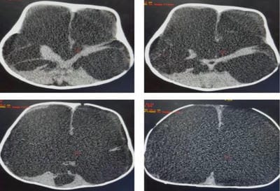
Figure 2: Axial non-contrast CT images of 2year old baby taken at multiple levels showing bilateral cerebral hemispheres being replaced by the fluid containing spaces of cerebrospinal fluid density. Normal posterior fossa, intact falx cerebri seen.

Discussion

The word hydranencephaly is a combination of hydrocephalous and anencephaly. It is a rare isolated anomaly occurring in less than 1 in 10000 births worldwide [5]. It is a rare encephalo-clastic anomaly characterized by the absence and replacement of the cerebral hemispheres with CSF [6]. There is the preservation of meninges, basal ganglia, pons, medulla, cerebellum and falx [7]. There is variable involvement of the inferior frontal, temporal and occipital lobes.


Falx cerebri is usually present but may be disrupted or absent with the septum pellucidum usually absent. The pathognomic feature of hydranencephaly is the preservation of brain stem and posterior fossa structures [8].

The etiology is usually unknown. Various causes have been stated like-

1. Infarction- is the most common mechanism. Bilateral occlusion of the internal carotid artery and in rare cases medial carotid artery usually between 8th and 12th weeks of gestation leads to the absence of structures perfused by ICA/MCA and the structures perfused by posterior cerebral artery and basilar artery are spared and hence are perfused.

2. Infection- intrauterine infections like TORCH may cause necrotizing vasculitis which in turn may lead to hydranencephaly.

3. Hypoxia- hypoxia in the fetus can cause defuse hypoxic-ischemic brain necrosis.

4. Leucomalacia- is an extreme form of leukomalacia that develops multiple cystic cavities that end up coalescing inside the cranial cavity.

5. Exposure to toxins- maternal exposure to smoking, cocaine, sodium valproate.

6. Multiple gestations- especially in twin pregnancy, the death of one twin leads to the development and accumulation of embolic material.

7. Rare conditions like factor 13 deficiency, intracerebral hemorrhage and fowler syndrome [9].

At birth in many cases, the baby may be normal [10]. An increase in head size is one of the early signs. Other signs are seizures, blindness, and global developmental delay [11]. The clinical presentation consists of a vegetative state with few reflexive functions like sucking, swallowing, crying and moving extremities, depending on the severity of the condition. The prognosis is poor with a usually fatal outcome during the first year [12]. The differential diagnosis of hydranencephaly includes other causes of enlarging head size in an infant like hydrocephalous, holoprosencephaly and prosencephalon which can be differentiated with imaging studies [13]. The most important imaging modalities for the diagnosis of hydranencephaly include a neuro-sonogram, CT scan brain, and MRI brain. In most cases, the CT scan brain shows the absence of most of the structures

in the supratentorial region, also there is the preservation of structures like falx cerebri, thalami, and basal ganglia. Features that differentiate hydrocephalous from hydranencephaly on CT scan brain is the presence of a thin, uniform rim of residual cerebral cortex in hydrocephalous which is lacking in hydranencephaly and also the visualization of 3rd ventricle in hydrocephalous which is lacking in hydranencephaly [14].

Treatment of hydranencephaly is mainly supportive and most children usually have a short life span [15].

Conclusion

Hydranencephaly is one of the cause of increasing head size in the new born and clinicians need to do early imaging studies to make a diagnosis and differentiate it from other common causes of increasing head size in children so that morbidities are reduced and their quality of life can be improved.

Reference

01. Cecchetto G, Milanese L, Giordano R, Viero A, Suma V, Manara R. Looking at the missing brain: hydranencephaly case series and literature review. Pediatr Neurol. 2013 Feb;48(2):152-8. doi: 10.1016/j.pediatrneurol.2012.10.009 [Crossref][PubMed][Google Scholar]

02. Tsai JD, Kuo HT, Chou IC. Hydranencephaly in neonates. Pediatr Neonatol. 2008 Aug;49(4):154-7. doi: 10.1016/S1875-9572(08)60032-7 [Crossref][PubMed][Google Scholar]

03. Malik AM, Ahmad M, Khan A, Ullah E. Hydranencephaly: a rare cause of delayed developmental milestones. BMJ Case Rep. 2013 Apr 30;2013:bcr2013009589. doi: 10.1136/bcr-2013-009589 [Crossref][PubMed][Google Scholar]

04. Pant S, Kaur G, De JK. Hydranencephaly. Kathmandu Univ Med J (KUMJ). 2010 Jan-Mar;8(29):83-6. doi: 10.3126/kumj.v8i1.3227 [Crossref][PubMed][Google Scholar]

05. Sultana, Z. , Kumari, A. and Pathan, H. , 2020. A Rare Case Of Hydranencephaly: A Case Report. International Journal of Anatomy and Research, 8(3.2), pp.7644-7648 [Crossref][PubMed][Google Scholar]


06. Azizah Hamida, Andi Arwan. Premature infant with Hydrancephaly: a first reported case in Indonesia. Asia Pacific Journal of Paediatrics and Child Health. 2020 Oct-Dec (3). [Crossref][PubMed][Google Scholar]

07. Greco F, Finocchiaro M, Pavone P, Trifiletti RR, Parano E. Hemihydranencephaly: case report and literature review. J Child Neurol. 2001 Mar;16(3):218-21. doi: 10.1177/088307380101600311 [Crossref][PubMed][Google Scholar]

08. Sepulveda W, Cortes-Yepes H, Wong AE, Dezerega V, Corral E, Malinger G. Prenatal sonography in hydranencephaly: findings during the early stages of disease. J Ultrasound Med. 2012 May;31(5):799-804. doi: 10.7863/jum.2012.31.5.799 [Crossref][PubMed][Google Scholar]

09. Pavone P, Praticò AD, Vitaliti G, Ruggieri M, Rizzo R, Parano E, et al. Hydranencephaly: cerebral spinal fluid instead of cerebral mantles. Ital J Pediatr. 2014 Oct 18;40:79. doi: 10.1186/s13052-014-0079-1 [Crossref][PubMed][Google Scholar]

10. Olowu JA, Lagunju IA, Tongo OO, Atalabi M. Intrauterine fetal death of one of twins, coexisting with hydranencephaly in the surviving co-twin: a case report. West Afr J Med. 2006 Jul-Sep;25(3):246-8. doi: 10.4314/wajm.v25i3.28288 [Crossref][PubMed][Google Scholar]

11. Adeloye A. Hydranencephaly in Malawian children. East Afr Med J. 2000 Jun;77(6):316-8. doi: 10.4314/eamj.v77i6.46641 [Crossref][PubMed][Google Scholar]

12. Khalid M, Khalid S, Zaheer S, Redhu N, Ekramullah. Hydranencephaly: a rare cause of an enlarging head size in an infant. N Am J Med Sci. 2012 Oct;4(10):520-2. doi: 10.4103/1947-2714.102015 [Crossref][PubMed][Google Scholar]

13. Kelly TG, Sharif UM, Southern JF, Gururajan K, Segall HD. An unusual case of hydranencephaly presenting with an anterior midline cyst, a posterior calcified mass, cerebellar hypoplasia and occlusion of the posterior cerebral arteries. Pediatr Radiol. 2011 Feb;41(2):274-7. doi: 10.1007/s00247-010-1894-1 [Crossref][PubMed][Google Scholar]

14. Daniyan, Olapeju Wunmi, et al. Hydranencephaly: A Cause of Increasing

Head Size in A Nigerian Neonate. " Archives of Clinical and Medical Case Reports 4. 3 (2020): 541-545. [Crossref][PubMed][Google Scholar]

15. Takada K, Shiota M, Ando M, Kimura M, Inoue K. Porencephaly and hydranencephaly: a neuropathological study of four autopsy cases. Brain Dev. 1989;11(1):51-6. doi: 10.1016/s0387-7604(89)80009-9 [Crossref][PubMed][Google Scholar]