E-ISSN:2349-3267
P-ISSN:2349-5499

Research Article

Blood transfusion

Pediatric Review - International Journal of Pediatric Research

2023 Volume 10 Number 1 January February
Publisherwww.medresearch.in

Prevalence, Pattern and Outcome of Blood Transfusion: A Private Paediatric Hospital Experience

A. West B.1*, Josephine E A.2
DOI: https://doi.org/10.17511/ijpr.2023.i01.01

1* Boma A. West, (FWACP), Department of Paediatrics and Child Health, Rivers State University Teaching Hospital, Nkpolu-Oroworukwo, Port Harcourt, Nigeria. 0000-0002-9849-1412

2 Aitafo Josephine E, (FWACP), Department of Paediatrics and Child Health, Rivers State University Teaching Hospital, Nkpolu-Oroworukwo, Port Harcourt, Nigeria. 0000-0003-3711-1495

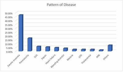
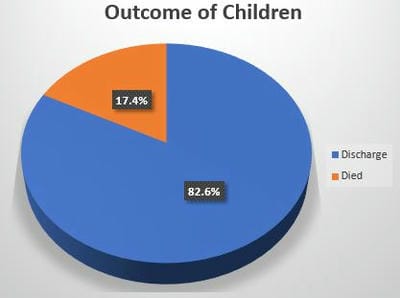
Introduction: Blood transfusion is an essential component of Pediatric care worldwide, especially in developing countries. Knowledge about its prevalence and pattern is vital in promoting its’ judicious use. Purpose: To ascertain the prevalence, pattern and outcome of blood transfusion in a private pediatric hospital in south-south Nigeria. Materials and Methods: A retrospective study was carried out over 1 year on children (0-17 years) admitted and transfused. Relevant data was retrieved from the hospitals’ Health Management System and analysed. Results: Of 1689 admissions, 69 were transfused giving a prevalence rate of 4.1% with male predominance (M: F ratio of 1.5:1). Eighty-eight blood transfusions were done giving a ratio of 1.3 transfusions/child. Transfused children were mainly 1–12 months of age with a mean age of 3.848 4.890 months. Most blood transfusions took place within the first 5 hours of prescription and occurred mostly at night. Sedimented cells were mainly used 49(71.0%). Nine(13.0%) children had blood transfusion reactions of which 5(60.0%) had a fever. The commonest diagnosis among children transfused were severe anaemia 61(46.9%), prematurity 22(16.9%), sickle cell anaemia 8(6.2%) and sepsis 7(5.4%). Twelve(17.4%) children transfused died. The age of children, the type of blood transfused and the number of transfusions carried out were significantly associated with the outcome of blood transfusion. Conclusion: The prevalence of blood transfusion was 4.1%. The commonest diagnosis was severe anaemia, prematurity, sickle cell anaemia and sepsis with a mortality rate of 17.4%. It is therefore important to equip private hospitals with functional blood bank services to reduce childhood morbidity and mortality.

Keywords: Blood transfusion, Outcome, Pattern, Prevalence

Corresponding Author How to Cite this Article To Browse
Boma A. West, (FWACP), Department of Paediatrics and Child Health, Rivers State University Teaching Hospital, Nkpolu-Oroworukwo, Port Harcourt, Nigeria.
Email:
Boma A. West, Aitafo Josephine E, Prevalence, Pattern and Outcome of Blood Transfusion: A Private Paediatric Hospital Experience. Pediatric Rev Int J Pediatr Res. 2023;10(1):1-9.
Available From
https://pediatrics.medresearch.in/index.php/ijpr/article/view/734

Manuscript Received Review Round 1 Review Round 2 Review Round 3 Accepted
2023-01-04 2023-01-06 2023-01-13 2023-01-20 2023-01-27
Conflict of Interest Funding Ethical Approval Plagiarism X-checker Note
Nil Nil Yes 16%

© 2023by Boma A. West, Aitafo Josephine Eand Published by Siddharth Health Research and Social Welfare Society. This is an Open Access article licensed under a Creative Commons Attribution 4.0 International License https://creativecommons.org/licenses/by/4.0/ unported [CC BY 4.0].

Introduction

Blood is a vital human tissue which when correctly, rationally and timely administered, saves lives, improves health conditions and enhances patients’ outcomes. [1] Blood transfusion aims at the replacement of the volume and specific constituents of blood which play specific roles in oxygen carriage, clotting and immunity. [2] It is an essential component of Paediatric care worldwide more so in developing countries. [3,4] That blood transfusion is still rampant in this part of the world might be due in part to the high prevalence of infectious diseases, malaria, malnutrition, sickle cell anaemia and late presentation to the hospitals. [3,5-7]

Varying transfusion rates have been reported in Nigeria, from as low as 2.7% in Ado Ekiti [5] southwest, 7% in Aba [3] southeast, 17.9% in Abakaliki, Ebonyi state [8] southeast, 31.2% at Sagamu [4] southwest and as high as 41.2% reported in Enugu,[9] southeast Nigeria. Various reasons have been proffered for this difference seen such as the level of care offered by the health facility (primary, secondary or tertiary care) and thus facilities available for transfusion, the age range of children recruited for the study and the prevalence of various diseases both communicable and non-communicable.[1-4,8,9]

The main recipients of blood transfusions are children, especially in resource-poor countries where they account for a high percentage of blood usage (16-67%) when compared with 5% reported in advanced countries.[1,10,11] Several authors have further reported that the majority of these transfusions were in children less than five years.[3,4,6,12] Common indications for blood transfusion in children include severe malaria, infections, sickle cell anaemia, malnutrition, prematurity, trauma and malignancies.[1-4,7,8,13] Several studies have reported severe malaria as the leading indication for transfusion in children in our environment.[3-5,14] There could be adverse reactions to blood transfusion which vary from simple febrile episodes to life-threatening episodes of haemolysis.[1,15]

Knowledge about the prevalence and pattern of blood transfusion is vital in promoting its’ judicious use, especially in resource-poor settings such as ours. To the best of our knowledge, this evaluation has not been carried out in any private

paediatric health facility in South-south Nigeria. This study was therefore carried out to ascertain the prevalence, pattern and outcome of blood transfusion in a privately owned paediatric hospital in south-south Nigeria. It would also contribute to the provision of baseline data for future reference.

Methods

This was a retrospective study involving all children admitted and transfused in a private paediatric hospital in Port Harcourt, Rivers State, over 1 year (from 1st of January 2021 to 31st of December,2021). Our study centre is a 38-bedded private hospital with a well-equipped neonatal unit, children’s ward, fully functional radiology unit and medical laboratory with attached blood bank services. It also has a fully functional theatre with an ultra-modern paediatric anaesthetic machine, piped oxygen supply and a paediatric ventilator. The age group seen was 0-17 years with an average monthly admission rate of 80-90 children per month. Its specialist staff strength included 6 paediatricians, a dermatologist, a paediatric surgeon, an ENT surgeon, a neurosurgeon, a burns and plastics surgeon, an orthopaedic surgeon, 2 anaesthetists, 2 radiologists, a radiographer as well as other support staff including nurses.

Data on all children admitted and transfused during the study period was retrieved from the admissions, theatre and blood bank records of the hospitals’ Health Management System. Information obtained included age, sex, diagnosis, indication for transfusion, number and type of transfusions, time of transfusion, time lag before transfusion, duration of admission, mode of payment of bills and outcome. Outcome measures included discharge, death, left against medical advice and referral.

Blood transfusion was prescribed for severe anaemia defined as packed cell volume (PCV) < 31% for newborns and PCV < 15% for all other children or otherwise higher PCV when a patient showed features of heart failure, [1,7] or severe haemorrhages, overwhelming septicaemia, severe neonatal jaundice etc. Transfusion for severe anaemia was with 15mls/kg of partially packed/sedimented cells because of a lack of facilities for packed red cells. For acute haemorrhage, bleeding disorders and sepsis, whole blood was used at 20mls/kg. Platelet or plasma concentrates were not used due to unavailability.


In almost all cases, blood for transfusion was sourced from voluntary non-renumerated donors recruited by patient relations. In some cases, blood used was obtained from the blood bank. Screening for HIV, HBV, HCV and VDRL was carried out routinely in our hospital blood bank on all potential donors and only those confirmed to be negative were allowed to donate. Double-volume exchange blood transfusion (EBT) was done in the newborns for cases of severe neonatal jaundice while single-volume EBT or top-up transfusions were done for severe anaemia and overwhelming septicaemia. Blood deemed fit for transfusion was properly grouped, cross-matched and labelled.

Data were recorded in an Excel spreadsheet and analysed using SPSS version 23. Results were presented as frequency, percentages, pie and bar charts. Test of association was done using the χ2 test and Fishers’ Exact test. Statistical significance was set at P value < 0.05 while results were reported as odds ratios at 95% confidence intervals.

Result

Demographic Characteristics
Of 1689 admissions, 69 children were transfused with blood giving a prevalence of blood transfusion of 4.1%. Eighty-eight blood transfusions were carried out on 69 children giving a ratio of 1.3 blood transfusions per child. Males predominated 41(59.4%) with an M: F ratio of 1.5:1. Majority of children transfused were 1 – 12 months of age 33(47.8%) with a mean age of 3.848 ± 4.890 months. The majority had a duration of stay of 1 – 7 days 37(53.6%) with a mean duration of stay of 7.026 ± 3.848 days. Most blood transfusions took place within the first 5 hours of prescription with the mean time lag before the 1st, 2nd and 3rd transfusions being 6.16 ± 3.085 hours, 6.55 ± 4.275 hours and 5.00 ± 4.583 hours respectively. Most children were transfused at night 53(60.2%) and paid out of pocket 45(65.2%). The mean pre-transfusion PCV was 27% ± 1.420 while the post-transfusion PCV was 38.753% ± 1.369. The mean rise in PCV after transfusion was 11.682% ± 1.761, Table I. 

The pattern of Blood Transfusion
The blood group of most of the children who had a blood transfusion and their donors were O Rhesus D positive, 28(43.8%) and 53(76.8%) respectively.

The majority of the blood donors were fathers 29(42.0%). Sedimented cells were majorly transfused 49(71.0%) and most children had a single blood transfusion 55(79.7%). Nine (13.0%) children had blood transfusion reactions of which 5(60.0%) had a fever and 4(40.0%) had skin rashes, Table II. 

Table I: Demographic Characteristics

Variables Frequency, n=69 (%)
Sex
 Male 41 (59.4)
 Female 28 (40.6)
Age (months)
 < 1 19 (27.5)
 1 – 12 33 (47.8)
 > 12 17 (24.6)
Duration of Stay (days)
 1 – 7 37 (53.6)
 > 7 32 (46.4)
Time lag before transfusion (hours)
 1 – 5 38 (55.1)
 ≥ 6 31 (44.9)
Time of transfusion (n=88)
 8.00am – 5.59pm 35 (39.8)
 6.00pm – 7.59pm 53 (60.2)
Payment Method
 Insurance 24 (34.8)
 Out of pocket 45 (65.2)

The pattern of Diagnosis Among Children who had Blood Transfusion
The commonest diagnosis among children transfused were severe anaemia 61(46.9%), prematurity 22(16.9%), sickle cell anaemia 8(6.2%) and sepsis 7(5.4%), Figure 1.

The outcome of Children who had Blood Transfusions
Of 69 children who received blood transfusion, 57(82.6%) were discharged home while 12(17.4%) died, Figure 2. 

Factors Associated with Blood Transfusion Outcome
The age of the children who had blood transfusions was significantly associated with the patient outcome, P value = 0.031, Table III. 

Association of Pattern of Blood Transfusion and Patient Outcome
There was a significant association between the type of blood transfused and the number of transfusions with the patient outcome (P value = < 0.001 and 0.001 respectively), Table IV. 


Association of Pattern of Disease and Patient Outcome
Amongst children who received blood transfusion, prematurity was significantly associated with patient outcome (P value = 0.007), Table V. 

Table II: Pattern of Blood Transfusion

Variable Frequency, n=69 (%)
Childs’ blood group
 A Rhesus D Positive 16 (25.0)
 A Rhesus D Negative 1 (1.6)
 B Rhesus D Positive 16 (25.0)
 O Rhesus D Positive 28 (43.8)
 O Rhesus D Negative 3 (4.7)
 Not recorded 5 (7.2)
Donors’ blood group
 A Rhesus D Positive 3 (4.7)
 B Rhesus D Positive 4 (5.8)
 O Rhesus D Positive 53 (76.8)
 O Rhesus D Negative 4 (6.3)
 Not recorded 5 (7.2)
Donors’ relationship to child
 Father 29 (42.0)
 Mother 9 (13.0)
 Parent’s relatives 14 (20.3)
 Parent’s friends 2 (2.9)
 Commercial 15 (21.7)
Type of blood transfused
 Sedimented cells 49 (71.0)
 Whole blood 17 (24.6)
 Both 3 (4.3)
Number of transfusions
 1 55 (79.7)
≥2 14 (20.3)
Presence of blood transfusion reaction(s)
 Yes 9 (13.0)
 No 60 (87.0)
Reactions observed
 Fever 5 (60.0)
 Skin Rash 4 (40.0)

SCA - Sickle cell anaemia, LPD – Lymphoproliferative diseases, NNJ – Neonatal jaundice

Figure 1: Pattern of Disease among Children who had Blood Transfusion

Table III: Factors Associated with Blood Transfusion Outcome

Variables Hospital Outcome Bivariate Test P value
Died, n=12(%) Discharged, n=57(%)
Sex      
Male 8 (66.7) 33 (57.9) 0.749
Female 4 (33.3) 24 (42.1)
Age (months)      
< 1 7 (58.3) 12 (21.1) 0.031*
1 – 12 3 (25.0) 30 (52.6)
> 12 2 (16.7) 15 (26.3)
Duration of Stay (days)      
1 – 7 3 (25.0) 34 (59.6) 0.053
> 7 9 (75.0) 23 (40.4)
Payment Method      
Insurance 5 (41.7) 19 (33.3) 0.740
Out of Pocket 7 (58.3) 38 (66.7)

*=Statistically significant

Table IV: Association of Pattern of Blood Transfusion and Patient Outcome

Variables Hospital Outcome Bivariate Test P value
Died, n=12 (%) Discharged, n=57 (%)
Child’s blood group      
A Rhesus D Positive 5 (50.0) 11 (20.4) 0.302
A Rhesus D Negative 0 (0.0) 1 (1.9)
B Rhesus D Positive 1 (10.0) 15 (27.8)
O Rhesus D Positive 3 (30.0) 25 (46.3)
O Rhesus D Negative 1 (10.0) 2 (3.7)
Donor’s blood group      
A Rhesus D Positive 1 (9.1) 2 (3.8) 0.155
B Rhesus D Positive 0 (0.0) 4 (7.5)
O Rhesus D Positive 8 (72.7) 45 (84.9)
O Rhesus D Negative 2 (18.2) 2 (3.8)
Donor’s relationship to child      
Father 3 (25.0) 26 (45.6) 0.322
Mother 1 (8.3) 8 (4.0)
Parent’s relative 5 (41.7) 11 (19.3)
Commercial 3 (25.0) 12 (21.1)
Type of blood transfused      
Sedimented cells 3 (25.0) 46 (80.7) < 0.001*
Whole blood 6 (50.0) 11 (19.3)
Both 3 (25.0) 0 (0.0)
Number of transfusions      
1 6 (50.0) 49 (86.0) 0.001*
≥2 6 (50.0) 8 (14.0)

*=Statistically significant


Figure 2: Outcome of Children who had Blood Transfusion 

Table V: Association of Pattern of Disease and Hospital Outcome

Variables Hospital Outcome Bivariate Test Fishers’ Exact
Died, n=12 (%) Discharged, n=57 (%)
Severe anaemia 11 (91.7) 50 (87.7) 1.000
Prematurity 8 (66.7) 14 (24.6) 0.007*
SCA 0 (0.0) 8 (14.0) 0.334
Septicaemia 3 (25.0) 4 (7.0) 0.095
Heart failure 1 (8.3) 5 (8.8) 1.000
Bleeding disorder 1 (8.3) 4 (7.0) 1.000
Severe malaria 1 (8.3) 2 (3.5) 1.000
Thalassemia 1 (8.3) 2 (3.5) 1.000
LPD 0 (0.0) 3 (5.3) 0.634
NNJ 1 (8.3) 1 (1.8) 0.320
Others 3 (25.0) 7 (12.3) 0.235

SCA- Sickle cell anaemia; LPD – Lymphoproliferative disorders; NNJ – Neonatal jaundice; *- Statistically significant

Discussion

The prevalence rate of blood transfusion in the present study of 4.1% was comparable with the 4.28% reported in Akwa Ibom, Nigeria [16] but lower than the 6.19%, 8.8%, 7.0% and 8.9% reported in China,[17] Yemen [1] and other parts of Nigeria[3,13] respectively. Much higher prevalence of 12.3%, 12.4%, 15.8% and 16% were documented in Ghana,[18] Brazil,[19] Benin city,[20] Nigeria and Tanzania [21] respectively. The low prevalence rate in the present study could be because it was carried out in a private health facility, unlike all other studies which were carried

out in tertiary health facilities with the possibility of a wider range of disease conditions. The higher prevalence rate in all other studies could also be attributed to the fact that more critically ill children are usually seen more in tertiary health institutions. Geographic locations and disease variations over time could also be responsible for the varied prevalence of blood transfusion.

Blood transfusion was carried out more in males in the present study with an M: F ratio of 1.5:1. This was similarly observed in other parts of Nigeria[4,16,22,23] as well as in China,[17] Yemen,[1] Ghana,[18] and England.[24] In contrast, a multi-institutional study in the USA[25] showed an almost equal Male: Female ratio whereas a retrospective study in Aba,[3] southeast Nigeria reported females being mostly transfused. This variation could be accounted for by the varying pattern of diseases and geographic locations as well as cultural differences as observed in Africa and other developing countries where the male gender is preferred and as such more likely to receive medical care as compared to their female counterparts.[26] Interestingly, morbidity and mortality rates were observed to be higher in males than females and thus, the possibility of the males being more likely to require blood transfusions.[27] This is because females have stronger humoral and cellular immune responses to infections and antigenic stimulations.[28]

Infants (0-12 months) constituted more than 3/4th of the children transfused in the present study. Of this percentage, about a quarter of the children were neonates ie 0-28 days old. Onyearugha et al [3] in southeast Nigeria also reported infants being the most transfused while Guo et al[17] in China reported children 0-28 days (neonates) as being the most transfused followed by children 1 month - 1 year. In addition, other studies in Nigeria, [4,23] and Yemen [1] documented most transfusions in the 1st 5 years of life. This is not surprising as this age group is usually most vulnerable to disease conditions because their immune systems are not fully developed as compared to the older age groups.[29] It is worthy of note that the younger the child, the more vulnerability.[29] Studies have shown that children < 5 years are mostly affected by severe anaemia and thus more likely to be transfused with blood products.[14,30] Contrary to the present study however, older children > 5-10 years were mostly transfused in a 6 months


retrospective study in the post-neonatal ward of a tertiary hospital in Sagamu, southwest Nigeria.[22] This difference could be due to the very short study period of 6 months as well as the fact that neonates were not included in this study.

There was a significant difference in the pre-transfusion and post-transfusion PCV in the present study while the mean rise in PCV after transfusion was 11.682% ± 1.761. The pre-transfusion PCV in the present study was much higher than the 15.2% ± 5.6 reported in Sagamu,[22] southwest Nigeria. This could be because neonates were part of the study population in the present study, unlike the latter in which they were excluded. The higher pre-transfusion PCV in the present study could also be explained by the fact that the definition of anaemia in newborns is much higher as compared with older children.[31] The mean rise in PCV after transfusion in the Sagamu study[22] was however higher than in the present study. This could be attributed to differences when post-transfusion PCV checks were carried out in the different studies as well as the type of blood transfused. In the present study, sedimented cells were transfused whereas, in the Sagamu[22] study, packed cells were used.

Close to half of the children transfused were of blood group O Rhesus D positive and more than three-quarters of the blood donated was of the same blood group. This was also reported by Al-Saqladi et al[1] in Yemen. This is not strange as 75% of Nigerians have this blood group type.[32] In addition, blood group AB Rhesus D negative is the rarest blood group accounting for 1% of the population in Nigeria [32] as also observed in the present study where no child was documented with this blood group.

Close to 3/4th of the blood transfused were sedimented red blood cells. This corroborates findings by Onyearugha et al[3] in Aba, southeast Nigeria. In contrast, red cell concentrate or packed cells were majorly used for transfusion in other parts of Nigeria,[16,22] USA,[25] India[33] and Yemen[1] whereas whole blood was the commonest blood type transfused in Benin city,[20] Nigeria and Cameroon.[23] The type of blood transfused is dependent on the pattern of disease or transfusion indications in the locality and the availability of the blood products as observed in the present study where severe anaemia was the commonest morbidity documented and as such

the need for sedimented cells. In the Yemen study,[1] red cell concentrates were mainly transfused because most children had chronic haemolytic anaemia. It is pertinent to note that platelet was not transfused in the present study due to its’ unavailability as also observed in Aba,[3] southeast Nigeria. Whole blood transfusion is therefore done in place of platelet concentrates in these centres.

Single transfusions accounted for more than three-quarters of transfusions in the present study. A similar pattern was reported in Yemen[1] and Benin city,[20] Nigeria. Ogunlesi et al[4] however in their 1-year cross-sectional descriptive study of children 0-15 years reported 68.4% of children received 2-4 sessions of blood transfusions while 31.6% had a single transfusion. This difference could be attributable to the varying degrees of anaemia as well as the morbidity pattern in the various geographic locations. It is pertinent to note that children who receive multiple blood transfusions are at risk of alloimmunization especially children with thalassemia and sickle cell anaemia.

There were blood transfusion reactions in 13% of children in the present study. This was comparable with the 11.6% in India[33] but higher than 4.4%, 3.8%, 0.9% and 0.17% documented in Yemen,[1] Brazil,[34], UK[24] and USA[35] respectively. The commonest transfusion reaction in the present study was fever as also documented in Yemen[1] and India[33] whereas allergic reaction was the commonest observed in Brazil.[34] The prevalence of blood transfusion reactions could be attributable to the availability of safe blood, the efficiency of blood transfusion services as well as the pattern of disease and their severity. It is pertinent to note that developed countries like the UK[24] and the USA[35] had the least prevalence of transfusion reactions at 0.9% and 0.17% respectively which could be because of the ready availability of safe blood and their more efficient blood transfusion services.

The disease pattern among children transfused in the present study was severe anaemia, prematurity, sickle cell anaemia and sepsis in descending order. Severe anaemia was also documented by Bateman et al[36] in their 6 monthly prospective multi-centre study of children < 18 months admitted in 30 paediatric intensive units as well as a Kenyan study.[37] Malaria was an uncommon disease condition among children transfused in


the present study as also observed in north-central Nigeria.[13] This was however not the case in other studies in Nigeria,[3,4,16,20,22] and Cameroon[23] where malaria was the commonest disease pattern among children transfused with blood. This difference could be attributed to the fact that the present study was in a private paediatric facility, unlike all the other studies which were carried out in tertiary health facilities thus the possibility of the different study populations with different socioeconomic status. Prematurity was the 2nd commonest morbidity pattern as neonates were included in the study, unlike many other studies in which this age group were excluded. In addition, varying methodologies and different diagnostic criteria could also account for these differences.

The mortality rate of 17.4% among transfused children in the present study was comparable with the 16% reported in Aba,[3] southeast Nigeria but higher than the 12.4%, 9%, 8.2% and 6.2% reported in Yemen,[1] Kenya,[38] Tanzania[21] and Benin city[20] southern Nigeria respectively. A much higher mortality rate of 21.5% was however documented in Ogun state,[4] southwest Nigeria. These variations in the mortality rates could be due to the varying underlying disease pattern, their severity, ready availability of blood products as well as availability of adequate manpower and infrastructural development in the health facilities including a functional blood bank. The high mortality in the present study is not surprising as severe anaemia being the commonest morbidity pattern is a significant cause of morbidity and mortality in children in Sub-Saharan Africa.[39] In addition, the lack of standard blood transfusion protocol in the country could also be responsible for the recorded high mortality rate in the present study.

Interestingly, the age of children was significantly associated with the outcome of blood transfusion as well as the type of blood transfused and the number of transfusions. In the present study, children < 1 month were significantly more likely to die as compared to other age groups. This is not surprising as newborn infants have much lower immunity as compared with older children. Children who received whole blood, those who received multiple blood transfusions and premature babies were significantly more likely to die. Thus, to reduce childhood morbidity and mortality, efficient blood transfusion services must be readily available in health facilities.

Conclusion

The prevalence of blood transfusion in a private paediatric health facility in southern Nigeria is low, being 4.1% with male predominance. The age group 0 – 12 months were mostly transfused while the disease pattern among children transfused was severe anaemia, prematurity, sickle cell anaemia and sepsis with a mortality rate of 17.4%. The age of children, the type of blood transfused and the number of transfusions carried out were significantly associated with the outcome of blood transfusion.

Blood transfusion is an important component of health care as it saves lives and improves patients’ outcomes. It is therefore imperative to equip even privately owned health facilities with manpower and infrastructure including a functional well-equipped blood bank to reduce childhood morbidity and mortality. The provision of national guidelines on standard transfusion protocol would also be beneficial. In addition, the reduction of the burden of the various underlying disease conditions will reduce the prevalence of blood transfusion.

Reference

1. Al-Saqladi AM, Albanna TA. A study of blood transfusion in paediatric patients at a teaching hospital, Aden, Yemen. Inter J Clin Transfus Med 2021; 9:1-9. http://doi.org/10.2147/IJCTM.S293720

2. Ogunlesi TA, Ogunfowora OB. Pattern and determinants of blood transfusion in a Nigerian neonatal unit. Niger J Clin Pract 2011; 14(3): 354-8. doi: 10.4103/1119-3077.86783.

3. Onyearugha CN, Okoronkwo NC, Onyemachi PE. Blood transfusion: Prevalence, indications and associated factors in children presenting to a tertiary health institution in southeast Nigeria. Amer J Paediatr 2021; 7(2): 53-6. Doi: 10.11648/j.ajp.20210702.13

4. Ogunlesi T, Fetuga B, Olowonyo M, Adekoya A, Adetola O, Ajetunmobi A. Severe childhood anaemia and blood Transfusion in a Nigerian secondary level facility. J Trop Pediatr, 2016; 62(2): 107-15. Doi: 10.1093/tropej/fmv083

5. Adegoke S, Ayansanwo A, Oluwayemi I, Okeniyi J. Determinants of mortality


in Nigerian children with severe anaemia. S Afr Med J 2010; 102(10): 807-10. doi: 10.7196/samj.5240

6. Muoneke VU, Ibekwe RC, Nebe-Agumadu HU, Ibe BC. Factors associated with mortality in under-five children with severe anaemia in Ebonyi, Nigeria. Indian Pediatr 2012; 49: 119-23. https://doi.org/10.1007/s13312-012-0026-4

7. West BA, Arthur A, Aitafo JE, Onunakwe H. Prevalence, associated factors and outcome of newborns with anaemia as seen in Rivers State University Teaching hospital, Nigeria. ACRI 2021; 21(4): 23-4. Doi: 10.9734/ACRI/2021/v21i430241

8. Muoneke V, Ibekwe R, Nebe-Agumadu H, Ibe B. Severe anaemia in under-five years children in Ebonyi State University Teaching Hospital, Abakaliki. Arch Dis Child 2010; 95: A100-01. http://dx.doi.org/10.1136/adc.2010.186338.218

9. Ughasoro MD, Ikefuna AN, Emodi IJ, Ibeziako SN, Nwose SO. Audit of blood transfusion practices in the paediatric medical ward of a tertiary hospital in southeast Nigeria. East Afr Med J. 2013; 90(1): 5-11.

10. Hassell OBI, Maitland K. Blood transfusion in resource-limited settings. In Hunter’s Tropical medicine. 9th ed. London: Magill-Maguire-Ryan-Solomon; 2012: 162-7.

11. McCormick M, Delaney M. Transfusion support: considerations in paediatric populations. Semin Hematol. 2020; 57(2): 65-72. Doi: 10.1053/j.seminhematol.2020.07.003

12. Shari CR, Sawe HR, Murray BL, Victor MG, Mfinanga JA, Runyon MS. Emergency blood transfusion practices among anaemic children presenting to an urban emergency department of a tertiary hospital in Tanzania. BMC Hematol. 2017; 17:19 doi: 10.1186/s12878-017-0091-y

13. Okpe ES, Abok II, Diala UM, Okolo SN, Joseph DE. Indications for Blood Transfusion among children in a tertiary hospital in north-central Nigeria. J Med Trop 2011; 13 (2): 95-7.

14. Austin NIR, Adikaibe EAB, Ethelbert OO, Chioma UE, Ekene NU. Prevalence and severity of malaria parasitaemia among children requiring emergency blood transfusion in a tertiary hospital in Imo state, Nigeria. Ann Med Health Sci Res 2014; 4(4): 619-23 doi: 10.4103/2141-9248.139349

15. Moncharmont P. Adverse transfusion reactions in transfused children. Transfus Clin Biol 2019; 26(4): 329-35. Doi: 10.1016/j.tracli.2019.08.002

16. Ino-Ekanem MB, Bassey EU. Overview of blood transfusion in a paediatric medical setting of a tertiary hospital in south-south Nigeria. Inter J Health Sci Res 2016; 6(7): 47-51

17. Guo K, Wang XY, Feng GS, Tian J, Zeng YP, Ma SX et al. The epidemiology of blood transfusion in hospitalized children: A national cross-sectional study in China. Transl Pediatr 2021; 10(4): 1055-61. doi: 10.21037/tp-20-464

18. Orish VN, Ilechie A, Combey T, Onyeabor OS, Okorie C, Sanyaolu AO. Evaluation of blood transfusions in anemic children in Effia Nkwanta Regional Hospital, Sekondi-Takoradi, Ghana. Am J Trop Med Hyg 2016; 94(3): 691-4. doi: 10:4269/ajtmh.15-0310

19. Goncalez TT, Sabino EC, Capuani L, Liu J, Wright DJ, Walsh JH et al. Blood transfusion utilization and recipient survival at Hospital das Clinicas in Sao Paulo, Brazil. Transfus 2012; 52(4): 729-38

20. Abhulimhen-Iyoha BI, Isreal-Aina TT. Emergency blood transfusion in children in a tertiary hospital in Nigeria: Indications, frequency and outcome. West Afr J Med 2018; 35(1): 20-4

21. Mosha D, Poulsen A, Reyburn H, Kituma E, Mtei F, Bygbjerg IC. Quality of paediatric blood transfusions in two district hospitals in Tanzania: A cross-sectional hospital-based study. BMC Pediatr 2009; 9: 51. doi:10.1186/1471-2431-1189-1151

22. Akodu SO, Adekambi AF, Gbadebo FA, Olanrewaju DM. Indications for blood and blood products transfusions among children in a semi-urban tertiary hospital in Nigeria. Ann Health Res 2020; 6(1): 36-43

23. Ayuketah PO, Tagny CT, Koki NP. Assessment of clinical blood transfusion practice in a pediatric tertiary hospital in Cameroon. J Blood Dis Ther 2019; 1: 101

24. Morley SL, Hudson CL, Llewelyn CA, Wells AW, Johnson AL, Williamson LM; EASTR study group. Transfusion in children: Epidemiology and 10-year survival of transfusion recipients. Transfus Med 2016; 26(2): 111-7. doi: 10.1111/tme.12283.


25. Slonim AD, Joseph JG, Turenne WM, Sharangpani A, Luban NL. Blood transfusions in children: A multi-institutional analysis of practices and complications. Transfus 2008; 48(1): 73-80, doi: 10.1111/j.1537-2995.2007.01484.x

26. Das Gupta M. Selective discrimination against female children in rural Punjab, India. Popul Dev Rev 1987; 13: 77-100

27. Lozano R, Naghavi M, Foreman KK, Lim S, Shibuya K, Aboyans V, et al. Global and regional mortality from 235 causes of death for 20 age groups in 1990 and 2010: A systematic analysis for the global burden of disease study 2010. Lancet 2012; 380(9859): 2095-128. DOI: https//doi.org/10.1016/S0140-6736(12)61728-0

28. Fish EN. The X-files in immunity: Sex-based differences predispose immune responses. Nat Rev Immunol 2008; 8: 737-44. https://doi.org/10.1038/nri2394

29. Why are Children more vulnerable to resistant infections? News and opinions 2019. Menu ReAct, reactgroup.org

30. World Health Organization. Hospital care for children: Guidelines for the management of common illnesses with limited resources. Geneva: WHO 2005. https://www.who.int/maternal_child_adolescent/documents/9241546700/en/

31. Lashkari C. Anaemia in newborns. News medical life sciences. news-medical.net/health. Cited 12/11/2022

32. Most common blood group in Nigeria + Rarest blood group in Nigeria. nimedhealth.com.ng

33. Ghataliya KJ, Kapadia JD, Desai MK, Mehariya KM, Rathod GH, Bhatnagar N, et al. Transfusion-related adverse reactions in pediatric and surgical patients at a tertiary care teaching hospital in India. Asian J Transfus Sci 2017; 11(2): 180-7, doi:10.4103/0973-6247.214348

34. Pedrosa AK, Pinto FJ, Lins LD, Deus GM. Blood transfusion reactions in children: Associated factors. J Pediatr (Rio J) 2013; 89(4): 400-6, doi:10.1016/j.jped.2012.12.009

35. Berg A, Courtney R, Krishnamurti L, Triulzi DJ, Yazer MH. Is intensive monitoring during the first transfusion in paediatric

patients necessary? Hematol 2014; 19(5): 304-8, doi:10.1179/16078454137.0000000122

36. Bateman ST, Lacroix J, Boren K, Forbes P, Barton R, Thomas NJ, et al. Anemia, blood loss and blood transfusions in North American children in the intensive care unit. Am J Respir Crit Care Med 2008; 178: 26-33. DOI: 10.1164/rccm.200711.16370C

37. Pedro R, Akoch S, Maitland R. Changing trends in blood transfusion in children and neonates admitted in Kilifi district hospital, Kenya. Malar J 2010;9: 300-14. https://doi.org/10.1186/1475-2875-9-307

38. Berkley JA, Ross A, Mwang I, Osier FHA, Mohammed M, Shebbe M, et al. Prognostic indicators of early and late death in children admitted to district hospital in Kenya: Cohort study. BMJ 2003; 326(7385): 361, doi:10.1136/bmj.326.7385.361

39. English M, Ahmed M, Ngando C, Berkley J, Ross A. Blood transfusion for severe anaemia in children in a Kenyan hospital. Lancet 2002; 359(9305): 494-5. doi. 10.1016/S0140-6736(02)07666-3