E-ISSN:2349-3267
P-ISSN:2349-5499

Research Article

Respiratory Distress

Pediatric Review - International Journal of Pediatric Research

2023 Volume 10 Number 2 March-April
Publisherwww.medresearch.in

Modality of Respiratory Support in Preterm Neonates with Respiratory Distress and its Outcome: Experience of a Tertiary Care Hospital in Bangladesh

Abdul Mannan M.1*, Mandal S.2, Rasel M.3, Biswas B.4, Mahabub M.5, Rita F.6, Afroze S.7
DOI: https://doi.org/10.17511/ijpr.2023.i02.02

1* Md. Abdul Mannan, Professor, Department of Neonatology, Bangabandhu Sheikh Mujib Medical University (BSMMU), Dhaka, Bangladesh.

2 Shimul Mandal, Registrar, Department of Neonatology, Cumilla Medical College Hospital, Cumilla, Bangladesh.

3 Mohammad Rasel, Resident, Year-5, Department of Neonatology, Bangabandhu Sheikh Mujib Medical University (BSMMU), Dhaka, Bangladesh.

4 Baisakhi Biswas, Master in Public Health (Community Medicine), National Institute of Preventive and Social Medicine (NIPSOM), Dhaka, Bangladesh.

5 Maria Mahabub, Resident, Year-5, Department of Neonatology, Bangabandhu Sheikh Mujib Medical University (BSMMU), Dhaka, Bangladesh.

6 Farhana Rita, Resident, Year-4, Department of Neonatology, Bangabandhu Sheikh Mujib Medical University (BSMMU), Dhaka, Bangladesh.

7 Sharmin Afroze, Assistant Professor, Neonatology Unit, Dr. M R Khan Shishu Hospital & Institute of Child Health, Dhaka, Bangladesh.

Background: Preterm neonates are at increased risk of developing respiratory distress after birth. In order to reduce preterm death and their morbidity, identification of etiology and optimum management of respiratory distress is crucial. Objective: To determine causes of respiratory distress, pattern of respiratory modality used and its outcome among admitted preterm newborns. Material and methods: This study included 142 preterm neonates admitted with respiratory distress and required respiratory support in the NICU of Bangabandhu Sheikh Mujib Medical University, during the period from January 2020 to December 2021. Causes of respiratory distress and pattern of respiratory support was observed. Morbidities and in-hospital mortality were compared between invasive and non-invasive support group in the study. Results: Mean gestational age among the studied neonates was 32.39 ± 2.46 weeks. Male was predominant (54.2%) and mean birth weight was 1631.44 ± 578.57 g. About 62% belonged to the gestational age within 28 - <34 weeks category. Respiratory distress syndrome (RDS) was the most common cause which was 50.0% following TTN 24.6%, congenital pneumonia 19.0% and PNA 6.3%. Sepsis, septic shock, DIC, NEC and in-hospital mortality, nasal trauma, ROP and IVH occurred significantly higher in the mechanical ventilator support group in comparison to the NIV support group and the p-value were <0.05. Conclusion: In our study Respiratory distress syndrome is the commonest cause of respiratory distress. Invasive ventilation was associated with significant morbidity and mortality in preterm neonates when compared with those required non-invasive ventilation.

Keywords: Neonates, Preterm, Mechanical ventilation, Non-invasive ventilation

Corresponding Author How to Cite this Article To Browse
Md. Abdul Mannan, Professor, Department of Neonatology, Bangabandhu Sheikh Mujib Medical University (BSMMU), Dhaka, , Bangladesh.
Email:
Md. Abdul Mannan, Shimul Mandal, Mohammad Rasel, Baisakhi Biswas, Maria Mahabub, Farhana Rita, Sharmin Afroze, Modality of Respiratory Support in Preterm Neonates with Respiratory Distress and its Outcome: Experience of a Tertiary Care Hospital in Bangladesh. Pediatric Rev Int J Pediatr Res. 2023;10(2):25-31.
Available From
https://pediatrics.medresearch.in/index.php/ijpr/article/view/743

Manuscript Received Review Round 1 Review Round 2 Review Round 3 Accepted
2023-03-29 2023-03-24 2023-03-31 2023-04-07 2023-04-14
Conflict of Interest Funding Ethical Approval Plagiarism X-checker Note
Nil Nil Yes 18%

© 2023by Md. Abdul Mannan, Shimul Mandal, Mohammad Rasel, Baisakhi Biswas, Maria Mahabub, Farhana Rita, Sharmin Afrozeand Published by Siddharth Health Research and Social Welfare Society. This is an Open Access article licensed under a Creative Commons Attribution 4.0 International License https://creativecommons.org/licenses/by/4.0/ unported [CC BY 4.0].

Introduction

The first 28 days of life, defined as neonatal period is the most vulnerable time for a child’s survival. Every year an estimated 4 million babies die in the first 4 weeks of life [1] accounting for more than half of the under-five child deaths in most regions of the world. [2] Almost all (99%) neonatal deaths are happening in developing countries. [3] Neonatal mortality rate is one of the indicators for measuring the health status of a nation. Though Bangladesh has achieved the MDG4 target for reduction of under 5 mortalities ahead of time but unfortunately neonatal mortality is still significantly high (30/1000 live birth) which accounts for 66.66% of all under 5 deaths. [4] Prematurity & its complications contributes to around 88% of all neonatal deaths in Bangladesh. [4] The mortality and morbidity of preterm neonates are significantly higher than those of full-term neonates because preterm neonates are more prone to develop respiratory failure. [5] The functional immaturity of their lung structure, can lead to impaired gas exchange and requires respiratory support.6 Respiratory distress is one of the most common problems that the neonates encounter within the first few days of life and require hospital admission. [2] Neonates with respiratory distress are 2-4 times more likely to die than neonates without respiratory distress. [7] In newborn, respiratory distress is recognized as one or more signs of increased work of breathing, such as tachypnea, nasal flaring, chest retractions, or grunting. [8] Respiratory distress syndrome (RDS) is by far the most common cause of respiratory distress in preterm infants (50.8%), followed by transient tachypnea of the newborn (4.3%) and pneumonia/sepsis (1.9%). [9] The optimal approach to the early respiratory management of the preterm infant remains controversial. [10-11] Most of the newborn with respiratory distress are managed by either non-invasive or invasive respiratory support. Noninvasive ventilation includes supplemental oxygen via a head box or by nasal cannula; continuous positive airway pressure (CPAP) via nasal prongs or mask; and heated humidified high-flow nasal cannula (HHHFNC). In order to reduce preterm death and their morbidity due to respiratory conditions, it is important to identify the etiology of respiratory distress, pattern of respiratory support they require and their outcomes. Currently there is scarcity of data in Bangladesh regarding the respiratory support

and outcome in preterm infants with respiratory distress. So, this study was conducted to reflect the short outcomes related to modality of respiratory support in preterm infants with respiratory distress for clinicians and other research workers.

Methodology

A retrospective descriptive study was conducted over a period of two years from January 2020 to December 2021 targeting all neonates admitted to neonatology department of Bangabandhu Sheikh Mujib Medical University. This department is a 31 bedded NICU equipped with advanced technologies for neonatal care. The unit provides level I, II and III neonatal care with multidisciplinary support. The NICU provides care for admissible inborn as well as critically sick newborns referred from different parts of the country.

Ethical approval was taken from Ethical Review Committee of the institution. All inborn preterm neonates having gestational age of <37 weeks and admitted with respiratory distress were included in the study. Neonates with incomplete data and or lethal congenital anomalies were excluded from the study. Data were collected from the neonatal admission, discharge and death registers. The register contained each neonate’s date of admission and discharge or death, sex, weight at admission or at birth, gestation age at birth, mode of delivery, duration of stay at the hospital, diagnosis and outcomes. The primary causes of admissions and deaths were defined as the underlying obstetric and neonatal factors or conditions, which resulted in the admission or death of the neonate. Standard definitions of the medical conditions were used for diagnosis.12 All medical and nursing staff working at the neonatal unit was oriented on recording of the neonatal admission, discharge and death registers, clinical guidelines of diagnosis and compilation of monthly summaries for presentation at monthly perinatal mortality meetings. Extracted data included: mode of delivery, multiple gestations, use of antenatal corticosteroid, gestational diabetic mellitus (GDM), Pregnancy induced hypertension (PIH), risk factor for sepsis, sex of the baby, gestational age (weeks), birth weight (g), APGAR score at 5th minutes, Silverman- Anderson score (SAS), neonatal resuscitation, surfactant administration, fetal growth at birth (SGA, AGA, LGA), respiratory distress, cause of respiratory distress, mode of respiratory support (NIV, MV) and days of hospitalization.


Neonates born at less than 37 completed weeks (less than 259 days) of gestation were termed as preterm and those having birth weight of < 2500 g were defined Low birth weight (LBW).12 Newborns was defined small for gestational age (SGA) if the birth weight less than the 10th percentile. Resuscitation was defined as need for intermittent positive pressure ventilation and/or cardiac compression and/or drug administration in the neonatal stabilization period. Respiratory distress in newborn was labelled when a baby had one or more signs of increased work of breathing, such as tachypnea, nasal flaring, chest retractions, or grunting.8 Severity of respiratory distress was categorized using Silverman-Anderson scoring system where Score ≥ 4 indicate clinical respiratory distress and score ≥ 7 indicate respiratory failure.13

Respiratory distress syndrome (RDS) was defined in neonates with increasing oxygen dependence during the first 24 h, typical radiological findings: like reduced air content, reticulo-granular pattern of the lungs, air bronchogram and/or white out lung. When newborns developed respiratory distress soon after birth and resolves within 18-24 hours of life with normal chest X-ray finding or show reduced translucency, infiltrates and hyperinflation of the lungs were labelled as Transient Tachypnea of newborn (TTN). Breathing pauses that last for > 20 seconds or for > 10 seconds if associated with bradycardia or oxygen desaturation was termed as apnea. Bronchopulmonary Dysplasia (BPD) was defined when a neonate was requiring oxygen at 36 weeks of post gestational age for babies born < 32 weeks of gestation or 28 days of age for neonates born ≥ 32 weeks of gestation or later.13

Maternal characteristics included as maternal age, maternal diseases like gestational and non-gestational diabetes mellitus, Pregnancy induced hypertension, infections and use of antenatal corticosteroids. Deliveries were categorized as vaginal or Cesarean Section (CS). The Data on respiratory support such as oxygen therapy administered through nasal-cannula, nasal continuous positive airways pressure (CPAP), heated humidified high flow nasal cannula (HHHFNC) and mechanical ventilation (MV) was collected.

When analyzing the data, for each neonate only the highest level of respiratory support was considered. The need for specific adjunctive therapy (surfactant

administration) and the short-term outcome including morbidities and in hospital mortality were recorded. Respiratory support was divided into two groups as Invasive (Mechanical ventilation) and noninvasive which included supplemental oxygen through nasal cannula, head box, High flow nasal cannula and continuous positive airway pressure (CPAP). All the variables and mortality were compared between the two groups

Statistical analysis: Data entry and analysis was carried out by using the Statistical Package of Social Science Software program (SPSS), version 22. Categorical variables were expressed in frequency and statistical analysis was done by Chi-Square test or Fisher exact test. Continuous variable was seen in mean ± SD and statistical analysis was done by student t-test. P value <0.05 was considered statistically significant.

Results

A Total of 733 patients were admitted in NICU during the study period from January 2020 to December 2021. Among them 50 neonates were excluded due to incomplete data and congenital malformations. Preterm babies were enrolled 234 in number (34.2%) and among them 142 (61%) were found to have respiratory distress requiring respiratory support as shown in figure-1.

pedia_743_01.JPG
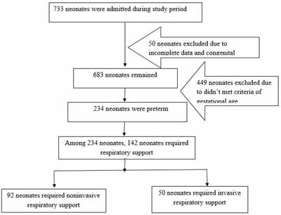
Figure 1: Flowchart of patient enrollment

Among 142 neonates, 92 required non-invasive respiratory support and remaining 50 patients needed invasive ventilator support along with non invasive ventilator support.

Baseline demographic and perinatal characteristics of the studied neonates were presented in Table-1


. Mean gestational age was 32.39 ± 2.46 weeks. Most of the newborns (62.0 %) belonged to the gestational age within 28 - <34 weeks category. Mean birth weight was 1631.44 ± 578.57 g among them very low birth weight infants were 41.5%.

Male out numbered female newborns (54.2% vs 45.8%). Total 09 (6.3%) infants had Apgar scores <7 at 5 minutes who required some degree of resuscitation just after birth. After admission 6 (4.2%) patients got single dose of surfactant.

Table-1: Baseline neonatal characteristics in studied neonates (N=142)

Parameter Value
Gestational age (weeks), Mean ±SD 32.39±2.46
Gestational age category, n (%)  
 <28 weeks 4 (2.8)
 28-<34 weeks 88 (62.0)
 34-<37 weeks 50 (35.2)
Birth weight (g), Mean ± SD 1631.44±578.57
Birth weight category, n (%)  
 <1000 g 13 (9.2)
 1000-1499 g 59 (41.5)
 1500-2499 g 57 (40.1)
 ≥2500 g 13 (9.2)
Sex of the baby, n (%)  
 Male 77 (54.2)
 Female 65 (45.8)
Multiple birth, n (%)  31 (21.8)
Neonatal resuscitation, n (%) 9 (6.3)
APGAR score at 5th minute  
≥7 137 (93.7)
<7 9 (6.3)
Silverman Anderson Score at randomization  
<4 22 (15.5)
4-7 120 (84.5)
>7 0 (0.0)
Surfactant administration, n (%) 6 (4.2)


Table-2: Baseline maternal characteristics in studied group (N=142)

Parameter Value
Consanguinity present, n (%) 2 (1.4)
Parity, n (%)  
 Primipara 46 (32.4)
 Multipara 96 (67.6)
Exposure to ACS, n (%)  
 Complete 24 (16.9)
 Incomplete 44 (31.0)
 None 74 (52.1)
Mode of delivery, n (%)  
 NVD 27 (19.0)
 LUCS 115 (81.0)
GDM, n (%) 43 (30.3)
PIH, n (%) 99 (69.7)
Risk factors for sepsis, n (%) 34 (23.9)

Regarding maternal characteristics (Table-2), most of the mother (67.6%) were multiparous and 52.1% of them did not receive even a single dose of antenatal corticosteroid. All of them were inborn and cesarean section was the mode of delivery for 81.0% of the enrolled neonates. Maternal hypertension and diabetes mellitus were present in 99 (69.7 %) and 43 (30.3%) of mother respectively. Maternal risk factors for sepsis were present in 34 (23.9%) of infants admitted to the NICU. 

Table-3: Primary disease requiring respiratory support (N=142)

Parameter Value
RDS, n (%) 71 (50.0)
Transient tachypnoea of newborn, n (%) 35 (24.6)
Congenital Pneumonia, n (%) 27 (19.0)
Perinatal asphyxia, n (%) 9 (6.3)
Meconium aspiration syndrome, n (%) 0 (0.0)

Regarding the respiratory morbidity (Table-3), respiratory distress syndrome (RDS) was the most common cause 71(50.0%) following TTN 35 (24.6%), congenital pneumonia 27 (19.0%) and PNA 9 (6.3%).

Table-4: Level of respiratory support (N=142)

Parameter Value
Non invasive support, n (%) 92 (64.8)
Invasive support, n (%) 50 (35.2)

Among total of 142 patients, 92 (64.8%) patients required NIV support including oxygen, CPAP or HFNC and 50 (35.2%) need Invasive support during the hospital course (Table-4).

Among the associated mortality and morbidity, sepsis, septic shock, DIC, NEC and in-hospital mortality occurred significantly higher in the invasive support group (IV) in comparison to the NIV support group and the p-value were <0.05 (Table-5).

Table-5: Associated mortality and morbidity of neonates who required respiratory support (N=142)

Parameters NIV group (n=92) IV group (n=50) P value
PDA, n (%) 17 (18.5) 13 (26.0) 0.39
Sepsis, n (%) 30 (32.6) 38 (76.0) <0.001
Septic Shock, n (%) 14 (15.2) 31 (62.0) <0.001
DIC, n (%) 5 (5.4) 16 (32.0) <0.001
AKI, n (%) 12 (13.0) 10 (20.0) 0.33
NEC, n (%) 7 (7.6) 14 (28.0) 0.001
In-hospital mortality, n (%) 7 (7.6) 30 (60.0) <0.001

Among the complication of respiratory support only nasal trauma, sepsis, ROP and IVH occurred significantly higher in the MV support group in comparison to the NIV support group and the p-value were <0.05 (Table-6). None of the infants in NIV support group developed pneumothorax but in MV support group 2 (4.0%) infants developed pneumothorax.

Table-6: Complication of Respiratory support modality in preterm neonates (N=142)

Parameters NIV group (n=92) IV group (n=50) P value
Nasal trauma, n (%) 9 (9.8) 16 (32.0) 0.001
Pneumothorax, n (%) 0 (0.0) 2 (4.0) 0.12
ROP, n (%) 5 (5.4) 9 (18.0) 0.016
BPD, n (%) 2 (2.2) 5 (10.0) 0.097
IVH, n (%) 0 (0.0) 7 (14.0) 0.001

Discussion

Preterm neonates are at high risk of developing respiratory distress in the immediate post-natal period. Optimum management of respiratory distress is mandatory to prevent mortality and morbidity. This retrospective study was done to evaluate the modality of respiratory support and outcomes in preterm neonates. Among total 234 inborn preterm neonates 142 (60.70%) needed respiratory support and were included in the study. In this study, mean gestational age of the studied neonates was 32.39 ± 2.46 weeks. Most of the newborns (62.0 %) belonged to the gestational age 28-<34 weeks category. Nemr CN, et al. showed 100 cases with gestational age ranging from 27 to 40 weeks having mean of 33.98 ± 3.44 weeks. Mean birth weight in our study was 1631.44 ± 578.57g which was comparable with the same study where mean birth weight was 1580 gm. [14] Male outnumbered female newborns (54.2% vs 45.8%). Similar finding was found by Nemr CH where sixty-three (63%) were boys and thirty-seven (37%) were girls and by Iqbal Q (60% males). [14-15] Regarding maternal characteristics, 52.1% did not receive even single dose of antenatal corticosteroid (ACS). All of them were inborn and cesarean section was the mode of delivery for 81.0% of the enrolled neonates. Maternal hypertension and diabetes mellitus were present in 69.7 % and 30.3% of neonates respectively. Maternal risk factors for sepsis were present in 23.9% of infants admitted to the NICU. In the study of Nemr CN, et al. 29% were multiple birth, LUCS 62% & maternal risk factor

for sepsis were 29%. In Lategan I, et al. 17.3% infants of < 34 weeks had optimal ANS, birth by Cesarean Section done in 60.3% of cases, maternal hypertension was present in 43.6% of mothers. [16] In our study, respiratory distress syndrome (RDS) was the most common cause 71(50.0%) following TTN 35 (24.6%), congenital pneumonia 27 (19.0%) and PNA 9 (6.3%) which were comparable with Nemr CN et al. RDS 50%, sepsis 38%, asphyxia 8% and pneumonia 4%.14 In this study, out of 142 patients, 64.8% patients required Non-invasive ventilator support including oxygen, CPAP or HFNC and 50 (35.2%) need Mechanical ventilator support during the hospital course. Nemr CN, et al. showed 72% of the studied patients underwent CPAP, 14% underwent oxygen support by nasal cannula and 14% underwent mechanical ventilation. [14] The need of invasive support was less than our study as they utilize non-invasive respiratory support as most widely used modality and practiced different ways of delivering CPAP. In the current study, non- invasive respiratory support was given by nasal cannula, head box, CPAP and invasive support by mechanical ventilation. We used nasal CPAP with preterm neonates with recurrent apneas or early features of RDS. Those who had a failure of nasal CPAP therapy or had respiratory acidosis were ventilated. This is similar to the results of Iqbal Q, et al. in which all the preterm neonates with gestational age < 32 weeks or recurrent apnea or early features of RDS were given nasal CPAP therapy, and those who had a failure of nasal CPAP therapy were ventilated. [15] Among the complications only nasal trauma, sepsis, ROP, and IVH occurred significantly higher in the MV support group in comparison to the NIV support group. In our study none of the infants in NIV support group developed pneumothorax but in MV support group 2 (4.0%) infants developed pneumothorax which is inconsistent with a previous study done by Nemr CN, et al. showed adverse effects occurred in 6 (43%) of them as: pneumothorax, ventilation acquired pneumonia and pulmonary hemorrhage. [14] Judicious ventilator strategies, proper sedation, timely extubation and readily available X-ray and surgical facilities will help to control these problems. Mortality among sick neonates in NICU is high, but mortality among mechanically ventilated neonates is even higher. In this study, mortality in ventilated neonates was 60%, which is comparable to mortality by Hossain et al. 70.6 %. [17]. Among the associated mortality and morbidity sepsis,


septic shock, DIC, NEC and in-hospital mortality occurred significantly higher in the Mechanical ventilator support group which is also evident in other studies. [18]

Conclusion

Respiratory distress syndrome is the commonest cause of respiratory distress.Two third of preterm newborns required respiratory support. Most common mode of respiratory support was non invasive mode in the form of supplemental oxygen, Heated humidified high flow nasal cannula and continuous positive airway pressure. Short term morbidities like nasal trauma, sepsis, septic shock, disseminated intravascular coagulation, necrotising enterocolitis and intraventricular haemorrhage were more common in newborns who required invasive respiratory support (p <0.05). Retinopathy of prematurity and mortality was significantly higher in invasive respiratory support group.

Limitations

  • Some patients were excluded due to incomplete data which might have a potential effect on outcome.
  • Some patients got 2 or more modalities of respiratory support during hospital course, althouth highest support was considerd for analysis.
  • Regarding patent ductus arterious, disseminated intravascular coagulation and intraventricular haemorrhage some patients were diagnosed clinically as investigation facility were not available for 24 hours period and also some patients died before confirming the diagnosis.

Reference

01. Lawn EJ, Cousens S, Zupan J, Lancet Neonatal Survival Steering Team. 4 million neonatal deaths. When? Where? Why? Lancet 2005 Mar;365(9462):891-900. . [Crossref][PubMed][Google Scholar]

02. Sweet LR, Keech C, Klein NP, Marshal HS, Tagbo BN, Quine D, et al. Respiratory distress in the neonate: Case definition and Guidelines for data collection, analysis, and presentation of maternal immunization safety data. Vaccine 2017; 35 (48 Pt

A): 6506-6517. . [Crossref][PubMed][Google Scholar]

03. Ayaz A, Saleem S. Neonatal mortality and prevalence of practices for newborn care in a Squatter Settlement of Karachi, Pakistan: A Cross-Sectional Study. PLOS One 2010; https://doi. org/10. 1371/journal.pone.0013783 [Crossref][PubMed][Google Scholar]

04. Bangladesh Demographic and Health Survey 2017–18. Dhaka and Rockville: National Institute of Population Research and Training and ICF; 2020. Available from: https:// dhsprogram. com/ pubs/ pdf/ FR344/ FR344. pdf [cited 2021 Aug 29] [Crossref][PubMed][Google Scholar]

05. Hong H, Li XX, Li J, Zhang ZQ. High-flow nasal cannula versus nasal continuous positive airway pressure for respiratory support in preterm infants: a meta-analysis of randomized controlled trials. The journal of maternal-fetal & neonatal medicine 2021;34 (2): 259–266. . [Crossref][PubMed][Google Scholar]

06. Raju, TNK. Developmental physiology of late and moderate prematurity. Seminars in Fetal and Neonatal Medicine 2012; 17(3), pp. 126–131. [Crossref][PubMed][Google Scholar]

07. Swarnkar K, Swarnkar M. Neonatal respiratory distress in early neonatal period and its outcome. Int J Biomed Adv Res. 2015; 6(9): 643–7. [Crossref][PubMed][Google Scholar]

08. Reuter S, Moser C, Baack M. Respiratory Distress in the Newborn. Pediatrics in Review 2014; 35(10), pp. 417–429. [Crossref][PubMed][Google Scholar]

09. Sharma, P, Poonia A, Bansal R. Comparison of efficacy of nasal continuous positive airway pressure and heated humidified high-flow nasal cannula as a primary mode of respiratory support in preterm infants. Journal of Clinical Neonatology 2019; 8(2), p. 102. [Crossref][PubMed][Google Scholar]

10. Jobe AH. Transition/adaptation in the delivery room and less RDS: “Don’t just do something, stand there!” Journal of Pediatrics 2005; Elsevier: pp. 284–286. . [Crossref][PubMed][Google Scholar]

11. Finer N. To intubate or not—that is the question: continuous positive airway pressure versus surfactant and extremely low birthweight infants. Archives of Disease in Childhood. Fetal and


Neonatal Edition 2006; 91(6): p. F392-F394 [Crossref][PubMed][Google Scholar]

12. Gomella TL, Cunningham DM, Eyal FG, editors. Neonatology: Management, Procedures, On-call Problems, Diseases and Drugs, Eighth edition. New York. Mc Graw Hill. Lange; 2020 [Crossref][PubMed][Google Scholar]

13. Hedstrom AB, Gove NE, Mayock DE, Batra M. Performance of the Silverman-Andersen Respiratory Severity Score in predicting PCO2 and respiratory support in newborns: a prospective cohort study. Journal of Perinatology 2018; 38: 505-511. . [Crossref][PubMed][Google Scholar]

14. Nemr CN, Bakheet MA, Hassan MA. Outcome of neonatal respiratory support modalities in Sohag University Hospital. The Egyptian Journal of Hospital Medicine. 2021; 82 (4): 778-784. [Crossref][PubMed][Google Scholar]

15. Iqbal Q, Younus MM, Ahmed A, Ahmad I, Iqbal J, Charoo BA, et al. Neonatal mechanical ventilation: Indications and outcomes. Indian Journal of Critical Care Medicine. 2015; 19 (9): 27-31. [Crossref][PubMed][Google Scholar]

16. Lategan I, Price C, Rhoda NR, Zar HJ, Tooke L. Respiratory interventions for preterm infants in LMICs: a prospective study from Cape Town, South Africa. Frontiers in Global Women’s Health. 2022; 3: 1-10. [Crossref][PubMed][Google Scholar]

17. Hossain MM, Shirin M, Mamun MA, Hasan MA, Shahidullah M. Predictors of mortality in ventilated neonates in Intensive Care Unit. Bangladesh J Child Health 2009; 33 (3):77-82. . [Crossref][PubMed][Google Scholar]

18. Basu B, Sinha SK, Basu T, Mahapatra TKS. Factors predicting mortality in newborn ventilation. Pediatr Pulmonol 2015; 50 (3):271-275. . [Crossref][PubMed][Google Scholar]