E-ISSN:2349-3267
P-ISSN:2349-5499

Case Report

Hypertrophic Cardiomyopathy

Pediatric Review - International Journal of Pediatric Research

2023 Volume 10 Number 3 May-June
Publisherwww.medresearch.in

Childhood Hypertrophic Cardiomyopathy with Double Chambered Right Ventricle: A rare coexistence

Mehrotra A.1*, Shakya U.2, Kacker S.3
DOI: https://doi.org/10.17511/ijpr.2023.i03.04

1* Akhil Mehrotra, Chief, Non Invasive Cardiologist Pediatric and Adult Cardiology, Prakash Heart Station, D-16 Nirala Nagar, Lucknow, Uttar Pradesh, India.

2 Ujala Shakya, Cardiac Technician, Prakash Heart Station, D-16 Nirala Nagar, Lucknow, Uttar Pradesh, India.

3 Shubham Kacker, Lead PMO, Tech Mahindra, New Delhi, India.

The prevalence of hypertrophic cardiomyopathy (HCM) is about 0.05-0.2% of the general population. The occurrence of HCM is a significant cause of sudden cardiac death in any age group and a cause of heart failure. The generally accepted definition of HCM is a disease state characterised by unexplained left ventricular (LV) hypertrophy without dilatation of ventricular chambers in the absence of another cardiac or systemic disease, which itself is capable of producing the amplitude of hypertrophy, caused by a genetic disorder in one of the at least ten genes that encode the proteins of the cardiac sarcomere. The prevalence of double-chambered right ventricle (DCRV) with childhood HCM is unknown and extremely rare. We are presenting here a unique coexistence of HCM with severe left ventricular outflow (LVOT) obstruction and DCRV in a 5year old male child.

Keywords: Hypertrophic Cardiomyopathy, Double Chambered Right Ventricle, left ventricular outflow obstruction, SAM

Corresponding Author How to Cite this Article To Browse
Akhil Mehrotra, Chief, Non Invasive Cardiologist Pediatric and Adult Cardiology, Prakash Heart Station, D-16 Nirala Nagar, Lucknow, Uttar Pradesh, India.
Email:
Akhil Mehrotra, Ujala Shakya, Shubham Kacker, Childhood Hypertrophic Cardiomyopathy with Double Chambered Right Ventricle: A rare coexistence. Pediatric Rev Int J Pediatr Res. 2023;10(3):51-56.
Available From
https://pediatrics.medresearch.in/index.php/ijpr/article/view/750

Manuscript Received Review Round 1 Review Round 2 Review Round 3 Accepted
2023-05-23 2023-05-25 2023-06-01 2023-06-08 2023-06-15
Conflict of Interest Funding Ethical Approval Plagiarism X-checker Note
Nil Nil Yes 18%

© 2023by Akhil Mehrotra, Ujala Shakya, Shubham Kackerand Published by Siddharth Health Research and Social Welfare Society. This is an Open Access article licensed under a Creative Commons Attribution 4.0 International License https://creativecommons.org/licenses/by/4.0/ unported [CC BY 4.0].

Introduction

HCM is the most common genetic disorder that can affect the left ventricular system with a prevalence of 0.05-0.2% of the general population. About 2/3 of patients experience left ventricular outflow obstruction known as hypertrophic obstructive cardiomyopathy (HOCM)[1] (Figure 1).

pedia_750_01.JPG
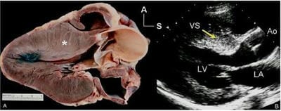
Figure 1: Long-axis anatomy typical of hypertrophic cardiomyopathy. A: Anatomic specimen shows a tremendous increase in left ventricular wall thickness, but the increase in myocardial mass is most prominently displayed in the ventricular septum (asterisk). B: Echocardiographic image displays similar anatomy. The increase in myocardial thickness is less prominent than in the anatomic example, but the basal septum (yellow arrow) is still asymmetrically thickened relative to the posterior left ventricular wall. A anterior; Ao, aorta; LA, left atrium; LV, left ventricle; S, superior; VS, ventricular septum.

DCRV is a rare congenital heart disease (CHD) characterized by anomalous muscle bundles (AMB) resulting in intra-cavitary obstruction in which the right ventricle is divided into a high-pressure chamber near the tricuspid valve and low-pressure chamber near the pulmonary valve (PV)[2] (Figure 2). Yamamoto et al first described HOCM in a DCRV patient with Noonan syndrome [3]

pedia_750_02.JPGFigure 2: Diagrammatic representation of Double Chambered Right Ventricle

Echocardiographic imaging plays an important role in the diagnosis of HCM with DCRV. Here we are presenting a case of HCM with severe LVOT obstruction associated with DCRV with moderate obstruction across the AMB, in a 5-year male child.

Case Report

A five-year male child presented to us for evaluation of a heart murmur. The parents enumerated the details of the patient and according to them, the child was asymptomatic since birth. On a routine check-up for a minor ailment, he was detected to be having a heart murmur. On clinical examination, the child was thin-built and healthy looking. The weight was 15 kg, height 60 cm, Pulse rate 76/min, respiratory rate 15/min, BP 80/50 right upper limb and SPO2 of 99% at room air. There was no evidence of any extracardiac musculoskeletal anomalies.

On cardiovascular examination, a Grade 3/6 ejection murmur was audible over the precordium best heard in the left second intercostal space, adjacent to the sternal edge. No radiation was detected in the carotids. All the peripheral arteries were normal and there was no radio-femoral delay.

pedia_750_03.JPG
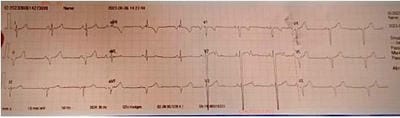
Figure 3: rS complexes from V1- V6, extreme left axis deviation, normal sinus rhythm

pedia_750_04.JPG
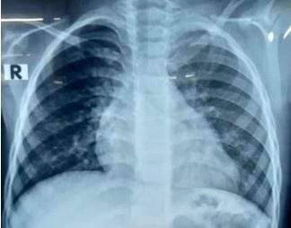
Figure 4: X-ray chest PA. There is cardiomegaly with normal pulmonary blood flow. 


ECG showed (Figure 3) rS compleves from V1-V6, extreme left axis deviation with normal sinus rhythm. X-ray Chest PA view (Figure 4) demonstrated mild cardiomegaly with normal pulmonary blood flow. Pathological reports were unremarkable.

Trans-thoracic echocardiography (TTE)

TTE was performed by the author in the supine and left lateral decubitus positions. Imaging was done from the subcostal, parasternal long axis (LX), parasternal short axis (SX), apical 4-chamber (4CH), apical 5-chamber (5CH) and suprasternal views.

There was levocardia, situs solitus, atrioventricular concordance, ventriculoatrial concordance, concordant d-subventricular loop, normally related great arteries and left aortic arch. Systemic and pulmonary venous drainage was normal.

In the LX views (Figure 5) asymmetrical septal hypotrophy is visualized with marked thickness of ventricular septum (Basal septum(D) 11.7 mm, Mid septum(D) 11.9 mm, Distal apical septum(D) 7.9 mm and left ventricular posterior wall(D) 5.2 mm,)

pedia_750_05.JPG

Figure 5: Parasternal LX View. There is the presence of marked thickness of the basal ventricular septum  

The interventricular septum and left ventricular posterior wall ratio was 2.2:1, consistent with hypertrophic cardiomyopathy.

M-mode echocardiography analysis at the level of the tip of mitral value demonstrated a classic systolic anterior motion (SAM) of the anterior mitral leaflet causing a severe LVOT obstruction (Figure 6).

pedia_750_06.JPG
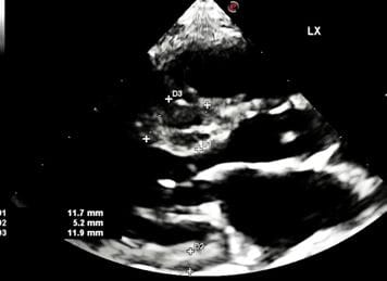
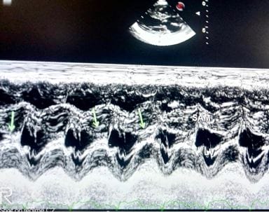
Figure 6: M-mode echocardiogram at the tip of the mitral valve shows that there is systolic anterior motion of mitral leaflets (SAM) towards the ventricular septum causing severe LVOT obstruction. 

pedia_750_07.JPG
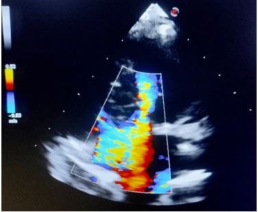
Figure 7: 5 CH View delineates a distinctive mosaic pattern in the LVOT, suggesting severe obstruction across LVOT.

pedia_750_08.JPG
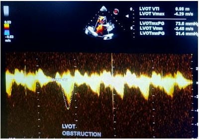
Figure 8: CW Doppler analysis of LVOT shows severe obstruction with a peak/ mean gradient across LVOT being 73.6/ 31.4 mmHg


In the 5CH view, a turbulent mosaic pattern is delineated in the LVOT, signifying a severe LVOT obstruction (Figure 7), with a peak/mean gradient of 73.6/31.4 mmHg (Figure 8). The marked thickness of anterolateral and posterolateral LV was also visualized.

Moreover in a modified 4CH view (Figure 9) a distinctive anomalous muscle bundle (AMB) was recognized in the middle of the right ventricular (RV) cavity, dividing the right ventricle into a proximal high-pressure chamber and a distal low-pressure chamber pressure.

pedia_750_09.JPG
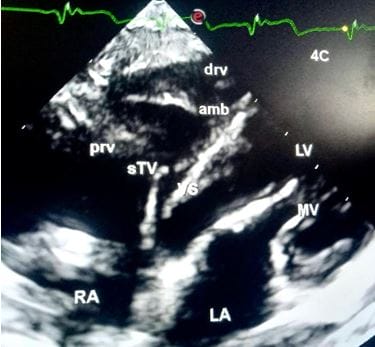
Figure 9. Modified 4 CH View demonstrates the characteristic presence of anomalous muscle bundle (AMB) near the apex of the right ventricle.drv, distal right ventricle, amb, anomalous muscle bundle, prv, proximal right ventricle, sTV, septal leaflet of Tricuspid Valve, VS, ventricular septum, RA, right atrium, LA, left atrium, MV, mitral valve, LV, left ventricle. 

pedia_750_10.JPG
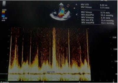
Figure 10: CW Doppler analysis across AMB shows a moderate grade of constriction with a peak/ mean gradient of 55.5/ 25.4 mmHg.

PG/MG across the AMB was 55.9/25.4 mmHg, suggesting a moderate grade of obstruction (Figure 10).

Anterior mitral leaflet (AML) and posterior mitral leaflet (PML) were large, thickened and redundant (Figure 11).

pedia_750_11.JPG
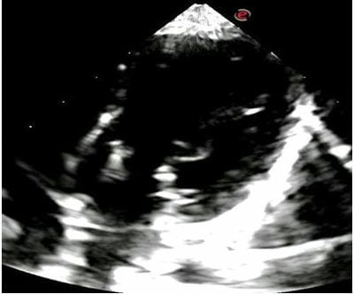
Figure 11: 4 CH View identifies large, thickened and redundant anterior and posterior mitral leaflets at the base of the left ventricular chamber. 

There was the presence of mild mitral regurgitation (MR), with an eccentric posterior jet measuring 1.34 sqcm. Nonetheless, there was mild dilatation of RV and LV size was small. Furthermore, there was normal biventricular systolic function and LVEF was 75%.

pedia_750_12.JPG
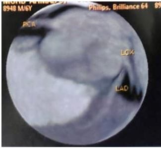
Figure 12: Coronary CT Angiography reveals normal coronary artery origins.


For ruling out the presence of anomalous coronary artery origins a 64-slice coronary CT angiography was performed and it demonstrated normal coronary anatomy (Figure 12).

Furthermore cardiac CT confirmed our echocardiographic diagnosis of asymmetrical septal hypertrophy and severe LVOT obstruction (Figure 13). The presence of HOCM with severe LVOT obstruction and additionally associated DCRV with moderate constriction obviates the need for corrective cardiac surgery and hence the patient was referred to a tertiary Paediatric Cardiovascular Institute.

pedia_750_13.JPG
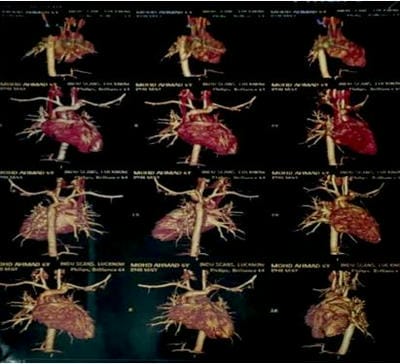
Figure 13: Cardiac CT suggestive of asymmetrical septal hypertrophy with remarkably thickened basal and mid-septum.

Discussion

The combination of HCM with DCRV is one of the extraordinary features of our case. A study showed that the annual incidence of cardiovascular mortality was 1-2% in adult patients with HCM [4] Sudden cardiac death (SCD) is one of the leading causes of death in patients with HCM, which is often correlated with lethal arrhythmias including ventricular tachycardia, ventricular fibrillation and a complete atrioventricular conduction block. Our patient is a 5year old child and is asymptomatic despite having severe LVOT obstruction at rest and to date had no symptoms of syncope/loss of consciousness or heart failure.

The double-chambered right ventricle is a rare disease in which the pressure

gradient across the obstruction frequently surpassed 20 mmHg [5] Approximately 80-90% of patients present with other cardiac anomalies, including VSD, ASD, subaortic stenosis, aortic valve regurgitation, PV stenosis, and so on[5]. However, DCRV associated with HCM is an extremely rare phenomenon. Several cases have earlier reported this distinctive combination [3, 6, 7]. Moreover, Ge et al. [8] described HCM in association with DCRV complicated by atrial flutter. Our patient had moderate DCRV which did not cause any haemodynamic or arrhythmic disturbances.

Conclusion

The coexistence of HCM with DCRV in childhood is an uncommon condition. Echocardiography and cardiac MRI are essential in the diagnosis of these anomalies. Due to the low risk of complications and a favourable long-term prognosis, surgery has proved to be fundamental in removing the obstruction. Clinicians should recognize early the risk factors of SCD in these patients and then stratify the risk to make the best decision in patients with HCM with DCRV.

Reference

01. Cheng Z, Fang T, Huang J, Guo Y, Alam M, Qian H. Hypertrophic Cardiomyopathy: from phenotype and pathogenesis to Treatment. Front Cardiovasc Med. (2021) 8:722340. [Crossref][PubMed][Google Scholar]

02. Loukas M, Housman B, Blaak C, Kralovic S, Tubbs RS. Anderson RH. Double-chambered right ventricle: a review. Cardiovasc Pathol. (2013) 22:417-23 [Crossref][PubMed][Google Scholar]

03. Yamamoto M, Takashio S, Nakashima N, Hanatani S, Arima Y, Sakamoto K, et al. Double-chambered right ventricle complicated by hypertrophic obstructive Cardiomyopathy diagnosed as Noonan syndrome. ESC Heart Fail. (2020) 7:721-6. [Crossref][PubMed][Google Scholar]

04. Elliott PM, Gimeno JR, Thaman R, Shah J, Ward D, Dickie S, et al. Historical trends in reported survival rates in patients with hypertrophic Cardiomyopathy. Heart. (2006) 92:758-91. [Crossref][PubMed][Google Scholar]

05. Hoffman P, Wojcik AW, Rozanski J, Siudalska H, Jakubowska E, Wlodarska EK, et al. The role of


echocardiography in diagnosing double chambered right ventricle in adults. Heart. (2004) 90:789-93. [Crossref][PubMed][Google Scholar]

06. Tyczynski P, Spiewak M, Chmielewski P, Kotlinski K, Deptuch T, Witkowski A, et al. Double chambered right ventricle in a patient with hypertrophic Cardiomyopathy. A unique coexistence. Kardiol Pol. (2021) 79: 891-2 [Crossref][PubMed][Google Scholar]

07. Said SM, Burkhart HM, Dearani JA, O’Leary PW, Ammash NM, Schaff HV. Outcomes of surgical repair of double-chambered right ventricle. Ann Thorac Surg. (2012) 93: 197-200. [Crossref][PubMed][Google Scholar]

08. Ge J, Hu T, Liu Y, Wang Q, Fan G, Liu C, Zhang J, Chen S, Maduray K, Zhang Y, Chen T, Zhong J. Case report: Double-chambered right ventricle diagnosed in a middle-aged female with hypertrophic Cardiomyopathy and atrial flutter: A rare case. Front Cardiovasc Med. 2022; 9: 937758. [Crossref][PubMed][Google Scholar]