E-ISSN:2349-3267
P-ISSN:2349-5499

Research Article

Plasma Lactate Dehydrogenase

Pediatric Review - International Journal of Pediatric Research

2023 Volume 10 Number 4 July-August
Publisherwww.medresearch.in

Association of Plasma Lactate Dehydrogenase concentration with Oxygen Dependence in Newborns with Respiratory Distress

Rahman M.1, Karmaker P.2, Rasel M.3, Tamima Nasrin U.4, Perveen J.5, Afreen S.6, Akhter S.7, Arif Hossain M.8, Abdul Mannan M.9*
DOI: https://doi.org/10.17511/ijpr.2023.i04.01

1 Mahbubur Rahman, Assistant Registrar, Department of Paediatrics, Shaheed M Monsur Ali Medical College & Hospital, Sirajgonj, Bangladesh.

2 Prohlad Karmaker, Assistant Registrar/Assistant Surgeon, Sheikh Sayera Khatun Medical College Hospital, Gopalgonj, Bangladesh.

3 Mohammad Rasel, Assistant Professor, Gonoshasthaya Samaj Vittik Medical College Savar, Savar, Dhaka, Bangladesh.

4 Ummey Tamima Nasrin, Assistant Surgeon, Department of Paediatrics, Upazilla Health Complex, Chandina, Cumilla, Bangladesh.

5 Jahanara Perveen, Consultant, Department of Paediatrics, OSD, DGHS, Dhaka, Bangladesh.

6 Shazia Afreen, Neonatologist, Department of Paediatrics, Kurmitola General Hospital, Dhaka Cantonment, Dhaka, Bangladesh.

7 Shamima Akhter, Medical Officer, Department of Paediatrics, Upazilla Health Complex, Homna, Cumilla, Bangladesh.

8 Md Arif Hossain, Medical Officer, Department of Neonatology, Bangabandhu Sheikh Mujib Medical University (BSMMU), Dhaka, Bangladesh.

9* Md. Abdul Mannan, Professor, Department of Neonatology, Bangabandhu Sheikh Mujib Medical University (BSMMU), Dhaka, Bangladesh.

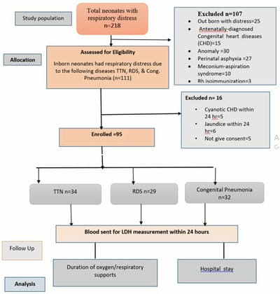
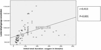
Background: Newborns with respiratory distress may need only supplemental oxygen, whereas those in advanced stages may require other respiratory supports like HHFNC, CPAP, and MV. Methods: This prospective observational study was carried out on 95 neonates, in the Department of Neonatology, BSMMU, Dhaka from July 2021 to June 2022. Enrolled infants were assigned into three groups: RDS group, TTN group, and congenital pneumonia group. LDH collection was done with all aseptic precautions within 24 hours of admission. Different modes of respiratory support were initiated in patients following NICU protocol according to their respiratory severity score. Respiratory supports were titrated according to the infant’s clinical condition, percent saturation of oxygen, and/or arterial blood gas analysis as per NICU protocol. Result: A total of 95 neonates were studied, mean values of LDH among the study groups were 755.64±222.70 u/l, 914.68±304.29 u/l and 742.81±284.70 u/l in TTN, RDS, and congenital pneumonia group respectively. High LDH levels were significantly associated with an increased need for oxygen support in the study group (P=0.04). In subgroup analysis showed high LDH was significantly associated with an increased need for oxygen support in TTN and congenital pneumonia group (P=0.024, P=0.001) respectively. Pearson correlation showed a positive correlation between LDH values and duration of oxygen supports (r=0.413, P=001). In this study, high LDH was also significantly associated with high respiratory support (p=0.001). There was no statistical association found between high LDH and hospital stay (P=0.165). Conclusion: High LDH was associated with the increased need for oxygen and advanced respiratory support among the baby with respiratory distress soon after birth.

Keywords: Plasma Lactate Dehydrogenase, Respiratory Distress, oxygen support

Corresponding Author How to Cite this Article To Browse
Md. Abdul Mannan, Professor, Department of Neonatology, Bangabandhu Sheikh Mujib Medical University (BSMMU), Dhaka, , Bangladesh.
Email:
Mahbubur Rahman, Prohlad Karmaker, Mohammad Rasel, Ummey Tamima Nasrin, Jahanara Perveen, Shazia Afreen, Shamima Akhter, Md Arif Hossain, Md. Abdul Mannan, Association of Plasma Lactate Dehydrogenase concentration with Oxygen Dependence in Newborns with Respiratory Distress. Pediatric Rev Int J Pediatr Res. 2023;10(4):69-77.
Available From
https://pediatrics.medresearch.in/index.php/ijpr/article/view/754

Manuscript Received Review Round 1 Review Round 2 Review Round 3 Accepted
2023-07-01 2023-07-04 2023-07-11 2023-07-18 2023-07-25
Conflict of Interest Funding Ethical Approval Plagiarism X-checker Note
Nil Nil Yes 18%

© 2023by Mahbubur Rahman, Prohlad Karmaker, Mohammad Rasel, Ummey Tamima Nasrin, Jahanara Perveen, Shazia Afreen, Shamima Akhter, Md Arif Hossain, Md. Abdul Mannanand Published by Siddharth Health Research and Social Welfare Society. This is an Open Access article licensed under a Creative Commons Attribution 4.0 International License https://creativecommons.org/licenses/by/4.0/ unported [CC BY 4.0].

Introduction

Approximately 30-40% of neonatal cases requiring hospitalization suffer from respiratory distress with high morbidity and mortality rates. Neonates with hypoxemia have a 3.1 times higher mortality rate[1]. Respiratory distress disorders are mostly related to hypoxia, due to delayed fluid transition, inefficient surfactant, tissue damage, etc [2]. Enzyme leakage as a result of hypoxia-ischaemia-induced cell damage in affected organs is well known[3]. Some markers which result from hypoxia-ischaemia induced cell damage in affected organs. Lactate dehydrogenase (LDH), lactate and also cystatin-C (Cys-C) are good predictors of hypoxia-ischaemia in the affected organs [4]. Lactate dehydrogenase (LDH) is found in most body tissues [5]. Lactate dehydrogenase (LDH) is an intracellular enzyme which converts pyruvic acid to lactic acid during the process of glycolysis [6]. It is released as a result of hypoxia and poor tissue perfusion [5]. Therefore, plasma LDH is also an indicator of body tissue hypoxia.[7]. LDH is one of these potential parameters presented in the literature as a possible indicator of lung damage [8]. High levels of LDH in the blood indicate tissue damage, so the LDH test is used to find out and monitor tissue damage, especially in respiratory diseases [9]. Emergency treatment in cases of neonatal respiratory distress is to reverse any hypoxia with supplemental oxygen and to prevent or reverse any respiratory acidosis by ensuring adequate ventilation of the lungs. This may require noninvasive respiratory support, such as continuous positive airway pressure (CPAP) or high-flow therapy [10]. A study by Ozkiraz et al., 2013, on respiratory distress in neonates also showed a relationship between LDH levels and the duration of supplying oxygen [7]. A study was done by Kamath et al., 2019, on prognosticating tool in NICU stay and oxygen dependence show LDH levels can be used as a prognosticating tool in predicting NICU stay and oxygen dependence.  Although some studies showed, there are relationships between LDH with a duration of 02 or respiratory supports and NICU stay but the evidence is still insufficient in our country. Therefore, this study aimed to find the association of LDH within 24 hours of life with oxygen dependence/duration of oxygen support of newborns with respiratory distress.


Methodology

This prospective observational study was conducted in the Department of Neonatology at Bangabandhu Sheikh Mujib Medical University (BSMMU), Shahbag, Dhaka over 12 months From July 2021 to June 2022. (Twelve months). Inborn babies (Term and Preterm) were admitted within 24 hours of birth having respiratory distress (due to Respiratory distress syndrome, Transient tachypnea of the newborn, and Congenital pneumonia) were included. Newborns delivered with major congenital anomalies, Respiratory distress due to Perinatal asphyxia, Meconium aspiration syndrome, Cyanotic congenital heart diseases, Hyper-bilirubinemia within 24 hours due to ABO or RH incompatibility and Death and DORB within 24 hours were excluded from this study. This study was approved by Institutional Review Board.

Study Procedure: Neonates with respiratory distress due to RDS, TTN, and Congenital pneumonia admitted to the NICU of BSMMU were enrolled in the study after getting informed consent from the parents. Neonates were transferred to the neonatal intensive care unit (NICU) after delivery room management. On the first day of hospitalization, details of maternal and perinatal medical history were taken and physical examination was done. Gestational age was calculated from the 1st day of the last menstrual period (LMP) in a mother with a previous regular menstrual cycle. In cases where LMP was not known, early obstetric ultrasonography was used to determine the gestational age. In cases where both were missing, gestational age assessment was made by using the New Ballard Score [11]. All required information for each neonate was recorded in a data collection form. The diagnosis of RDS, TTN, and congenital pneumonia was done according to history, symptoms, and laboratory evaluation including chest radiograph.

Enrolled infants were assigned into three groups RDS group, TTN group, and congenital pneumonia group according to diagnosis.  Oxygen dependence of neonates was assessed when they had clinical distress (tachypnea, chest retraction, cyanosis), poor oxygen saturation (<90%) or abnormal blood gas (Po2<60 mmHg) and got oxygen support equal and more than 120 hours.


For this oxygen dependence, different modes of respiratory support were initiated in patients according to their respiratory severity score (Downe’s score/Silverman score where needed).

Supplemental oxygen support through the nasal catheter with a flow rate of 1-2 L/min was initiated if the score was 1-2 and Oxygen through the head box with a flow rate of 3-5 L/min was initiated if the score was 3. An infant with Downe’s score of 4 or more was managed with non-invasive ventilation support through a heated humidified high-flow nasal cannula (HHHFNC) with an initial set-up of flow 5L/min and FiO₂ 0.4 or continuous positive airway pressure (CPAP) with an initial set-up of Pressure 5 cm of H₂O, flow 5 L/min and FiO₂ 0.4 as per NICU protocol. If the baby not improved after the height set up of CPAP (Term pressure 9 cm of H20 and Fio2 >.6, Preterm pressure 8 cm of H20 and Fio2 >.6) then the baby is put on mechanical ventilation.

Respiratory support was titrated according to the infant’s clinical condition, percent saturation of oxygen and arterial blood gas as per NICU protocol. Arterial Blood Gas analysis measurement was usually done 1-2 hours later after putting on HHHFNC or CPAP. The subsequent ABG was done after a change in parameter following clinical deterioration. An arterial blood sample of 0.3 ml (30 units) was drawn from the radial or posterior tibial artery and measured in NICU within 3-5 minutes. ABG machine Model OPTI CC TS blood gas analyzer was used in this study.

Blood was collected for plasma LDH measurement in a heparinized tube with all aseptic precautions within 24 hours of admission and 2.0 millilitres of sample were sent for investigation in the laboratory within 15 to 30 minutes. Then samples were collected by a lab technician and waited for 15 minutes, and then centrifuged @ 3500 -4000 RPM for 10 minutes. LD reagent is used to measure lactate dehydrogenase activity by an enzymatic rate method in BECKMAN COULTER (made in the USA) /ATELLICA machine (made in Germany). The SYNCHRON System(s) performs all calculations internally to produce the final reported result. Results were expressed as U/L (Normal values of LDH 330-700 U/L). A cut-off value of more than 700 U/L was used to define the high LDH level. Then newborns were classified into the normal LDH group and the high LDH group.

The goal of providing respiratory support was to maintain a stable respiratory condition (respiratory rate 30 – 60 breaths/minute, adequate chest expansion, no chest retraction, no grunting or cyanosis), SpO₂ ranging between 90% to 95% and an acceptable arterial blood gas (Pᴴ 7.35 to 7.45, PCO₂ 35 to 45 mm Hg, PO₂ 50 to 80 mm Hg and HCO₃ 24 ± 2 mmol/L). Respiratory support was gradually reduced with improvement in the respiratory condition and then stopped. The baby’s respiratory status was monitored for 6 hours till the withdrawal of respiratory support.

The duration of total respiratory supports (means the duration of oxygen or respiratory supports got by babies from first initiation of respiratory support to the baby just after admission up to first weaning from respiratory supports or continuation of initial oxygen support up to baby took DORB or death when babies were getting oxygen supports). The total duration of respiratory support was calculated in hours. Data about the respiratory support were obtained until the requirement of oxygen. The total duration of NICU stay was taken for the studied neonates. The diagnoses, set by the resident doctor on duty with the help of a consultant, then registered when the patient was discharged, died or took DORB during a hospital stay.

Data analysis: After collection data were entered into a personal computer and edited, analyzed, and plotted in graphs and tables whenever necessary. Data were analyzed using the statistical package for social sciences (SPSS) version 22. Quantitative data were expressed as mean and standard deviation and categorical data were presented as frequency and percentage. All quantitative variables were compared by unpaired t-test; categorical variables were compared by Chi-square test or Fisher’s exact test. The correlation of continuous variables was assessed by the Pearson correlation coefficient. A p-value less than 0.05 was considered statistically significant.

Flow chart of study procedure:

A total of 218 newborns developed respiratory distress after birth during the study period. According to the inclusion and exclusion criteria, 111 babies were eligible for this study. Among 111 cases, 16 were excluded from this study due to 5 Cyanotic congenital heart diseases, 6 developed jaundice within 24 hours and 5 cases were excluded


as parents did not give consent. So, the remaining 95 neonates were enrolled. Then three (3) groups were divided into Transient tachypnea of newborn (TTN=34), Respiratory distress syndrome (RDS=29) and Congenital pneumonia group (32) based on history, clinical exam and laboratory investigation including chest radiograph. The primary outcome was followed up, analyzed, and compared among the three groups. Outcomes were available for all the patients until discharge or death from the neonatal intensive care unit (NICU)

pedia_754_01.JPG

Figure: Flow Chart of Patient Enrollment and their Outcome.

Results

Table 1 shows baseline characteristics of neonates in the studied group, mean gestational age was 36.58±1.01weeks, 33.12±1.71weeks and 35.90±1.42weeks in the TTN, RDS and congenital pneumonia group respectively. Mean birth weights among the study groups were 36.58±1.01 gram, 33.12±1.71 gram and 35.90±1.42 gm in TTN, RDS and congenital pneumonia groups respectively. Among the study groups, LUCS was done in 67 (70.50%) and NVD was done in 28 (29.4%) respectively, most of them were singleton (87.3%). All of them were inborn. The mean respiratory severity score was 2.55±.89, 3.89±.30 and 2.62±.87 in TTN, RDS and congenital pneumonia groups respectively.


Table 1: Baseline characteristics of neonates in the studied group (N= 95)

Parameter TTN (n=34) RDS (n=29) Congenital pneumonia (n=32)
Gestational age in weeks (mean± SD) 36.58±1.01    
Gestational age category (weeks) (%)
(<34 wk) 4(13.8%) 20(69%) 5(17.2%)
(≥34 wk) 30(45%) 9(13.6%) 27(40.9%)
Birth weight (gm) (mean ± SD) 2659.70±404.87 1621.65±390.75 2328.90±373.42
Birth weight category, n (%)
(<1500 g) 2(11.1%) 13(72.2%) 3(16.7%)
(≥1500g) 32(41.6%) 16(20.8%) 29(37.7%)
Mode of delivery, n (%)
NVD 15(44.2%) 5(11.3%) 8(25%)
LUCS 19(55.8%) 24(82.7%) 24(75%)
APGAR score  at 1 min 6.91±0.28 6.89±0.30 6.96±0.17
APGAR score  at 5 min 7.11±0.32 7.3±0.48 7.2±0.25
Number of Gestation, n (%)
Single 32(94.1%) 22(75.8%) 29(90.6%)
Multiple 2(5.9%) 7(24.2%) 3(9.4%)
Gender of the baby, n (%)
Male 24(70.6%) 16(55.2%) 21(65.6%)
Female 10(29.4%) 13(44.8%) 11(33.7%)
Respiratory Severity Score, (Mean ± SD) 2.55±.89 3.89±.30 2.62±.87
Respiratory severity score category, n (%)
< 4 27(79.4%) 3(10.3%) 24(75%)
≥ 4 7(20.6%) 26(89.7%) 8(25%)

Categorical data are presented as number and frequency. Quantitative data are presented as mean ±SD , SD: Standard Deviation,, NVD- normal vaginal delivery, LUCS- lower uterine caesarian section. 

Table 2: Baseline maternal characteristics in the studied group (N=95)

Parameter TTN (n=34) RDS (n=29) Congenital Pneumonia(n=32)
Exposure to ACS, n (%)      
None 32(94.1%) 9(31%) 23(71.9%)
Incomplete 0(0.0%) 8(27.6%) 4(12.5%)
Complete 2(5.9%) 12(41.4%) 5(15.6%)
PROM>18hr n (%) 0(00%) 6(6.3%) 16(16.8%)
UTI n (%) 1(1.1%) 1(1.1%) 13(13.7%)
Asthma, n (%) 2(2.1%) 0(00%) 1(1.1)
Hypertension, n (%) 6(6.3%) 13(13.7%) 10(10.5%)
Gestational diabetes mellitus, n (%) 5(5.3%) 5(5.3%) 8(8.4%)

Categorical data are presented as number and frequency. Quantitative data are presented as mean ±SD, ACS- antenatal corticosteroid, GDM- gestational diabetes mellitus, HTN- hypertension.

Regarding maternal characteristics (Table 3), most of the mothers did not receive even a single dose of antenatal corticosteroid. But In subgroup analysis, antenatal corticosteroids got more in RDS groups than in other groups. Premature rupture of the membrane (>18hr) and UTI are mostly found in the congenital pneumonia group.

Table 3: LDH   laboratory values of neonates in the studied group (N= 95)

Parameter TTN (n=34) RDS (n=29) Congenital pneumonia (n=32)
LDH concentration(u/L) Mean ± SD   755.64±222.7   914.68±304.29   742.81±284.70
LDH concentration (u/L) Category (%)
Normal values (330-700 u/l) 15(35.7%) 9(21%) 18(42.9%)
High values (700-2000 u/l) 19(35.8%) 20 (37.7%) 14(26.4%)

Categorical data are presented as number and frequency. Quantitative data are presented as mean ±SD, LDH= Lactate Dehydrogenase concentration. 

Table 3: Laboratory values of LDH in neonate in studied groups, mean values among the study groups were 755.64±222.70 u/l, 914.68±304.29 u/l and 742.81±284.70 u/l in TTN, RDS and congenital pneumonia group respectively.

Table: 4: LDH values and their relation with duration of oxygen support in the studied group (N=95)

Oxygen dependence (≥ 120 hours) Normal LDH n=42 High LDH n= 53 p-value
Total, n (%)
< 120 hours 34(55.7%) 27(44.3%) 0.004s
≥ 120 hours 8(23.5%) 26((76.5%)
TTN, n (%)      
< 120 hours 15(53.6%) 13(46.4%) 0.024s
≥ 120 hours 0(00) 6(100%)
RDS, n (%)
< 120 hours 1(11.1%) 8(88.9%) 0.201ns
≥ 120 hours 8(40%) 12(60%)
Congenital pneumonia, n (%)
< 120 hours 18(75%) 6(25%) 0.001ns
≥ 120 hours 0(00%) 8(100%)

Statistical test: Fisher‘s Exact test, Chi-square test. S=significant ns= not significant.

Table: 4: showed among the study population, babies got more than 120 hours of oxygen support in the high LDH group (76.5%) than the normal LDH group (23.5%). There were significant differences found between both groups (P=0.004). Subgroup analysis showed there was a significant difference found in the duration of oxygen support of more than 120 hours in TTN and congenital pneumonia groups. P-values showed statistical differences in TTN (P= <0.05) and congenital congenital pneumonia (P=0.001) groups respectively.

pedia_754_02.JPG

Figure 3: Pearson Correlation of LDH concentration with an initial total duration of oxygen  (02)  in the studied group

The figure showed there is a significant positive correlation found in LDH concentration with an initial total duration of oxygen (P=0.001), (r=0.413).

Table: 5: LDH values and their relation with different mode respiratory supports in the studied group (95)

Modes of Respiratory Supports Normal LDH n=42 High LDH n= 53 p-value
Total, n (%)
Only Supplemental O2 32(60.4%) 21(39.6%) 0.001s
Respiratory supports 10(23.8%) 32(76.19%)
TTN, n (%)
Only Supplemental O2 15(53.6%) 13(46.4%) 0.02s
Respiratory supports 0(00) 6(100%)
RDS, n (%)
Only Supplemental O2 0(00) 1(100%) 1.00ns
Respiratory supports 9(32.1%) 19(67.9%)
Congenital pneumonia, n (%)
Only Supplemental O2 17(70.8%) 7(29.2%) 0.010s
Respiratory supports 1(12.5%) 7(87.5%)

Statistical test:  Fisher‘s Exact test, Chi-square test. S=significant ns= not significant


Table 5: showed among the study population out of 95 babies 42 (44.21%) babies got Respiratory support (all) more in the high LDH group (76.19%) than in normal LDH groups (23.8%) and there were significant differences found in between both groups (P=0.004). Subgroup analysis showed there were significant differences found in Respiratory supports in TTN(P=0.024)  and congenital pneumonia groups(P=0.010). 

Table: 6: LDH values with a prolonged hospital stay

Hospital stay Normal LDH (n=42) High LDH (n= 53) p-value
Total, n (%) Prolong hospital   stay
Yes 21(38.2%) 34(61.8%) 0.165ns
No 21(52.5%) 19(47.5%)
TTN, n (%) Prolong hospital  stay
Yes 3(27.3%) 8(72.7%) 0.271ns
No 12(52.2%) 11(47.8%)
RDS, n (%) Prolong hospital stay
Yes 8(34.8%) 15(65.2%) 0.633ns
No 1(16.7%) 5(83.3%)
Congenital pneumonia, n (%) Prolong hospital stay
Yes 10(47.6%) 11(52.4%)   0.266ns
No 8(72.7%) 3(27.3%)

Statistical test: Fisher‘s Exact test, Chi-square test. S=significant ns= not significant

Table 6: showed among the study population, babies got prolonged hospital stays in the high LDH group (61.8%) but there was no significant difference found between both groups (P=0.165). Subgroup analysis showed there were no significant differences found in prolonged hospital stay in TTN, congenital pneumonia and RDS  groups. P-values showed no statistical difference in TTN (P=0.271), congenital pneumonia (P=0.266) and RDS group(P=0.633).

Discussion

In this study, the mean gestational age were 36.58±1.01weeks, 33.12±1.71weeks and 35.90±1.42weeks in the TTN, RDS and congenital pneumonia group respectively, which is lower than previous studies done by Elfarargy et al. [8] (where mean gestational age were 37.1 ± 1.1 and 35.9 ± 1.3 in TTN and RDS group respectively) and S. Costa et al.,2012 (where mean gestational age were 38 weeks and 38 weeks in TTN and Pneumonia group respectively). A study was done in Germany where the mean gestational

age (RDS group) were 33.4±1.8 week [12] which is similar to my study (RDS group). Another study was done in Bangladesh where the mean gestational age for Pneumonia was 33±3.9 weeks [13] which is close to my study. A possible explanation is, the main bulk of our babies usually come from the Feto-maternal medicine department, they deal with critical deliveries and complicated pregnancies which ultimately leads to more preterm delivery.

In the present study, the mean birth weight was 2659.70±404.87g, and 1621.65±390.75g in TTN and RDS respectively which is lower than the previous study done by Lee M, et al., 2021 where the mean birth weight was 3288±484g, 3051 ± 210 g in TTN and  RDS group respectively. A Study was done in Portugal, where the mean birth weight in pneumonia was also higher (3400g) [14] than my study (2328.90±373.42g). Another study was done in Bangladesh by Mannan. A et al.[12] where the mean birth weight for Pneumonia was 2392±85g which is similar to my study(2328.90±373.42g). This difference can be explained by the recruitment of more very preterm and lower birth weight babies in this study.

The mode of delivery of most of the newborns was LUCS (70%) in this study which is higher than the previous study(58.9%) done by Lee M, et al [15]. Another study was done in Korea, where 62% of delivery occurred by Caesarean section [15] which is also lower than my study(70%). In this study, this higher percentage of the LUCS may be explained by the fact that this study was conducted in a tertiary care hospital, where most of the complicated pregnancies are dealt by the Feto-maternal medicine unit, with necessitating LUCS. In this study, most of the babies were male (64.21%) which is close to the previous study where the percentage of males is 62.9% [16].

In the present study, Mean LDH values among the study groups were 755.64±222.70 U/L,914.68 ±304.29U/L in TTN and RDS groups respectively which is much lower than previous study done by Elfarargy et al.[8], where mean LDH values were 820 ± 121 U/L,1112 ± 162 U/L, in TTN and RDS groups respectively. An et al. [14], and   Lee M, et al [15], showed high LDH values in both TTN and RDS groups than my studyA study was done in Romania, where mean LDH values in COVID-19 pneumonia in newborns were 546.17 ± 131.83 U/l [17], which is much lower than the


present study (742.81±284.70 U/L). This low mean LDH level in my study can be explained by the recruitment of more very preterm babies.

In our study, preterm (759±215 U/l) had lower levels of LDH values than term (862±349U/L) which is similar to a previous study done by Van Anh et al [14], where  LDH levels were higher in the full-term infants(751 U/l) than in the preterm infants(594 U/l). In another study done in Vietnam, mean plasma LDH levels were also lower in preterm infants [18]. This lower level of LDH might be either due to a physiological low level of LDH and glutamic-oxaloacetic transaminase and glutamic-pyruvic transaminase activity in preterm infants or decreased release of intra-cellular LDH caused by incomplete cell metabolism in response to hypoxia.

There was a paucity of data regarding mean LDH values of congenital pneumonia in other literature. But in some studies in infants, mean LDH values in pneumonia were much lower than my study (742.81±284.70 u/l). A study done in China showed mean LDH values in pneumonia were 415.00 u/L [19]. But in another study done by Kyoung Min Ann.et al. [7], where serum LDH levels (952 土 142 u/l) in pneumonia is more than that of my study.

This prospective observational study revealed some intriguing results. In this study, those newborns who needed oxygen support for 120 hours (5 days) or more had significantly high LDH(P=0.004) levels which is similar to previous studies done by Lee M, et al. [15], Ozkiraz, et al.[3]. A study done by Ozkiraz, et al. [3], showed a significant relationship between prolonged oxygen supplement(>72hr) and high LDH (750 U/l cut-off point) levels, with a specificity of 90.6%, a sensitivity of 47.6%, and an area under the curve of 73.3%.

In Subgroup analysis, the statistically significant association of high LDH with increased oxygen support (more than 120 hours) was found in TTN (P=0.02) and congenital pneumonia (P=0.001) groups which is similar to a previous study done by Lee M, et al. [15], wherein TTN groups average 5.8 days of oxygen support was needed. In the study done by An et al [14] in Korea, a significant association was found between high LDH with duration of oxygen support(P=0.013). Another study done in India showed an association between high LDH and an increased need for oxygen support (20).

In this study, the mean duration of oxygen support in the RDS group (9 days) is not significantly associated with High LDH (P=0.201) which is different from other studies like Lee M, et al. [15] (P=0.01). This can be explained by premature weaning of oxygen in about 9(37.5%) babies in the RDS group, this is due to either death or Left Against Medical Advice (LAMA). That’s why it could not reflect the actual average duration of oxygen support in the RDS group. Another possible explanation of this fact is the recruitment of more very preterm babies who needed more oxygen support but had decreased release of LDH for prematurity due to physiologically low levels of LDH or decreased release of intra-cellular LDH caused by incomplete cell metabolism in response to hypoxia.

In this study, about 55% baby got only supplemental oxygen and 44% baby got respiratory supports group where the mean LDH value are 699.u/l and 964.42u/l respectively which is close to similar to a previous study done by Ozkiraz et al [3]. Where mean LDH values in supplemental and respiratory supports group were 753±300 and 891±350 u/l.  A study done by Van Anh et al. [14], showed those babies requiring CPAP had high LDH (903u/l) than babies requiring no CPAP support (719 u/l). Elfarargy et al.[8], showed an association of high LDH with advanced respiratory supports. So the studies following my study suggest that increased LDH is associated with an increased need for respiratory support.

In this study, the mean duration of hospital stay is 9.1 days. High LDH is found to be associated with prolonged hospital stays which are following the result of previous studies done by Lee M, et al. [15], Liu Y et al. [18], and Kamath, M. et al [19]. But there was no significant association between high LDH and a prolonged hospital stay which is similar to the previous study done by Kamath, M.  et al., [19] where the p-value was found 0.889.

In subgroup analysis showed that the mean duration of hospital stay was 6.6 days in TTN and 13 days in the RDS group which is lower than the previous study done by Lee M, et al.[15], where the mean duration in RDS ( 15.1 days) and in TTN ( 9.1 days) were statistically significant which is in contrast to the result of the current study.

In my study, Pearson co-relation showed the association of high LDH values with


increased duration of oxygen supports (r=0.413, P=0.001) which is similar to a study done by Ozkiraz et al. [3], where a significant correlation was found between LDH values and duration of oxygen supports (r=0.43, P=0.001). Reference regarding the association of LDH and congenital pneumonia in the neonatal period is still lacking as there is no published study as yet in this field.

Measurement of the LDH is a low-cost test in most laboratories. So, LDH can act as an early predictor of the severity of the disease condition, especially respiratory diseases. LDH may be useful for clinicians at first-level hospitals for decision-making to refer the respiratory distressed newborn to the secondary or tertiary level neonatal intensive care unit before the clinical situation is worsened.

Limitations

1. A single sample was taken.

2. Sometimes LDH sample collection and sending to the laboratory was not possible in mid-night admitted babies due to my unavailability, for this I missed some study populations.

Conclusion

  • High LDH was associated with the increased need for oxygen and advanced respiratory support among the baby with respiratory distress soon after birth.
  • Also, this study showed an association between high LDH with an increased need for the duration of oxygen support in TTN and the congenital pneumonia group.

Recommendation:

1. LDH may be used as a predictor to assess the severity of the disease condition, especially respiratory diseases.

2. Need multicenter, large-scale study for further evaluation.

Abbreviation

BSID: Bayley scales of infant and toddler development

BSMMU: Bangabandhu Sheikh Mujib Medical University

IRB: Institutional Review Board

NICU: Neonatal intensive care unit

PNA: Perinatal asphyxia

ACS: Antenatal corticosteroid

BSMMU: Bangabandhu Sheikh Mujib Medical University

CPAP: Continuous positive airway pressure

FiO₂ : Fraction of inspired oxygen

GDM: Gestational diabetes mellitus

HHHFNC: Heated humidified high flow nasal cannula

HTN: Hypertension

LPT: Late preterm

LUCS: Lower segment caesarean section

PPV: Positive Pressure ventilation

SpO₂: Percent saturation of oxygen

SPSS: Statistical Package for Social Sciences

Reference

01. Rusmawati A, Haksari E, Naning R. Downes score as a clinical assessment for hypoxemia in neonates with respiratory distress. The Indonesian Journal of Paediatrics and Neonatal medicine. 2016; 48(6):342. [Crossref][PubMed][Google Scholar]

02. Kommawar, A. ‘Study of respiratory distress in newborn’, International Journal of Contemporary Pediatrics. 2017; 4(2): p. 490. [Crossref][PubMed][Google Scholar]

03. Ozkiraz S, Gokmen Z, Boke SB, Kilicdag H, Ozel D,and Sert A. Lactate and lactate dehydrogenase in predicting the severity of transient tachypnea of the newborn’, Journal of Maternal-Fetal and Neonatal Medicine. 2013; 26(12): 1245–1248. . [Crossref][PubMed][Google Scholar]

04. El Farargy, M. S. and Soliman, N. A. .Early Predictors of Transient Tachypnea of Newborn. Journal of Molecular Biomarkers & Diagnosis.2017; 08(02): 8–11 [Crossref][PubMed][Google Scholar]

05. Jahan, R. Diagnostic accuracy of lactate dehydrogenase for diagnosis of perinatal asphyxia in neonates with non-reactive CTG’, Pakistan Journal of Medical and Health Sciences. 2019; 13(2): pp. 458–460. [Crossref][PubMed][Google Scholar]


06. Dave, A. , Maru, L. and Jain, A. LDH (Lactate Dehydrogenase): A Biochemical Marker for the Prediction of Adverse Outcomes in Pre-eclampsia and Eclampsia’, Journal of Obstetrics and Gynecology of India. 2016; 66(1): 23–29 [Crossref][PubMed][Google Scholar]

07. Anh, T. N. Van and Hoang, H. H. The relationship between the plasma lactate dehydrogenase and severe conditions in newborn infant: A prospective study’, Pediatrics & Therapeutics.2018; 08 [Crossref][PubMed][Google Scholar]

08. Elfarargy, M. S. and Al-ashmawy, G. M. Novel predictor markers for early differentiation between transient tachypnea of newborn and respiratory distress syndrome in neonates. International Journal of Immunopathology and Pharmacology. 2021; 35:1-10 [Crossref][PubMed][Google Scholar]

09. Gallacher, D. J. , Hart, K. and Kotecha, S. 27064402.2016; 12(1): pp. 30–42 [Crossref][PubMed][Google Scholar]

10. Ballard JL, Khoury JC, Wedig K, Wang L, Eilers-Walsman BL, and Lipp R. New Ballard Score, expanded to include extremely premature infants. The Journal of Pediatrics. 1991; 119( 3): 417–423. [Crossref][PubMed][Google Scholar]

11. Lackmann, G. M. Influence of neonatal idiopathic respiratory distress syndrome on serum enzyme activities in premature healthy and asphyxiated newborns. American Journal of Perinatology. 1996; 13(6): pp. 329–334 [Crossref][PubMed][Google Scholar]

12. Mannan, M. Neonatal Pneumonia in NICU of a Tertiary Care Center’, Bangladesh Journal of Child Health. 2018; 42(3): pp. 112–117. [Crossref][PubMed][Google Scholar]

13. Costa, S. et al. Transient tachypnea of the newborn and congenital pneumonia: A comparative study’, Journal of Maternal-Fetal and Neonatal Medicine. 2012; 25(7): pp. 992–994 [Crossref][PubMed][Google Scholar]

14. An, Y. S. Serum Enzymes in Predicting Transient Tachypnea of Newborn and Respiratory Distress Syndrome’, Korean Journal of Perinatology. 2014; 25(4): p. 284 [Crossref][PubMed][Google Scholar]

15. Lee, M , Lee, N , Bae, H, Han,M, Park,H , Byun, Y and Kim,C. Using lactate

dehydrogenase to predict the severity of respiratory distress in term newborn infants with no perinatal asphyxia. Turkish Journal of Pediatrics. 2021; 63(3): 393–403. [Crossref][PubMed][Google Scholar]

16. Stoicescu, E. R. The Assessment of COVID-19 Pneumonia in Neonates: Observed by Lung Ultrasound Technique and Correlated with Biomarkers and Symptoms’, Journal of Clinical Medicine. 2022; 11(12). [Crossref][PubMed][Google Scholar]

17. Karlsson, M. Lactate dehydrogenase as an indicator of severe illness in neonatal intensive care patients: A longitudinal cohort study’, Acta Paediatrica, International Journal of Paediatrics. 2012; 101(12): pp. 1225–1231. [Crossref][PubMed][Google Scholar]

18. Liu Y, Shen Y,and Wei B. The Clinical Risk Factors of Adenovirus Pneumonia in Children Based on the Logistic Regression Model: Correlation with Lactate Dehydrogenase. Int J Clin Pract. 2022; Vol. 2022,1-3 [Crossref][PubMed][Google Scholar]

19. Kamath, M. K. Asha,M, Saihari, B and Sneha, M. Lactate Dehydrogenase as a Prognosticating Tool in Predicting NICU Stay and Oxygen Dependence in Meconium Stained Amniotic Fluid Neonates’, Journal of Clinical and Diagnostic Research. 2019; 11–13 [Crossref][PubMed][Google Scholar]