
Giant Congenital Melanocytic Nevi: A Case Report and Review of Literature
Keywords:
Congenital, Giant, Melanoma, Nevi, Neurocutaneous
Abstract
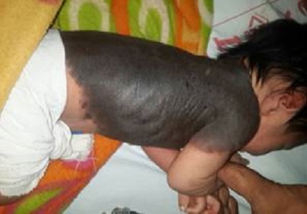
The congenital melanocytic nevi which are formed by the overgrowth of melanocytes occur in about 1% of the newborns. Giant Congenital Melanocytic Nevi (GCMN) which are of sizes larger than 20 cm diameter are rare and they occur in 1/500,000 newborns. Primary diagnosis of congenital giant nevus is clinical. Here, we report a case of full-term infant born with extensive black patch having smooth surfaces, irregular margins, and covering 45% of the skin surface.
Downloads
Download data is not yet available.
References
1. Walton RG, Jacobs AH, Cox AJ. Pigmented lesions in newborn infants. Br J Dermatol. 1976 Oct; 95 (4): 389–96. [PubMed]
2. Caro William A. Tumours skin, In: Dermatology, Edited by Moschello SL, Pillshury DM, Hurley HJ, WB Saunders Company, Philadelphia, 1978, p. 1323-1407. [PubMed]
3. Viana AC, Gontijo B, Bittencourt FV. Giant congenital melanocytic nevus. An Bras Dermatol. 2013 Nov-Dec;88(6):863-78. doi: 10. 1590/ abd 1806-4841. 20132233. [PubMed]
4. Rhodes AR. Melanocytic precursors of cutaneous melanoma. Estimated risks and guidelines for management. Med Clin North Am.1986 Jan;70(1):3–37. [PubMed]
5. Rhodes AR, Weinstock MA, Fitzpatrick TB, Mihm MC Jr, Sober AJ. Risk factors for cutaneous melanoma. A practical method of recognizing predisposed individuals. JAMA. 1987 Dec 4;258(21):3146-54. [PubMed]
6. Reed WB, Becker SW Sr, Becker SW Jr, Nickel WR. Giant pigmented nevi, melanoma, and leptomeningeal melanocytosis : A clinical and histopathological study.Arch Dermatol. 1965 Feb; 91:100–19. [PubMed]
7.Rhodes AR, Melski JW. Small congenital nevocellular nevi and the risk of cutaneous melanoma. J Pediatr. 1982 Feb; 100(2): 219-24. [PubMed]
8. Tannous ZS, Mihm MC Jr, Sober AJ, Duncan LM. Congenital melanocytic nevi: clinical and histopathologic features, risk of melanoma, and clinical management. J Am Acad Dermatol. 2005 Feb; 52(2): 197-203. [PubMed]
9. Iconomou T, Michelow BJ, Zuker RM. Tissue expansion in the pediatric patient. Ann Plast Surg. 1993 Aug; 31(2):134–40. [PubMed]
10. Kinsler V, Bulstrode N. The role of surgery in the management of congenital melanocytic naevi in children: a perspective from Great Ormond Street Hospital. J Plast Reconstr Aesthet Surg. 2009 May; 62 (5):595-601. doi: 10.1016/j.bjps.2008.12.016. Epub 2009 Feb 25. [PubMed]
11. Krengel S, Hauschild A, Schäfer T. Melanoma risk in congenital melanocytic naevi: a systematic review. Br J Dermatol. 2006 July; 155(1):1-8. [PubMed]
CITATION
DOI: 10.17511/ijpr.2016.i07.02
Published: 2016-07-31
How to Cite
Dr. Saurabh Piparsania, Dr. Saurabh Singh, Dr. Shubhangi Mahashabde, & Dr. Swati Raipurkar. (2016). Giant Congenital Melanocytic Nevi: A Case Report and Review of Literature. Pediatric Review: International Journal of Pediatric Research, 3(7), 479-481. https://doi.org/10.17511/ijpr.2016.i07.02
Issue
Section
Case Report
Copyright (c) 2016 Author (s). Published by Siddharth Health Research and Social Welfare Society

This work is licensed under a Creative Commons Attribution 4.0 International License.

 OAI - Open Archives Initiative