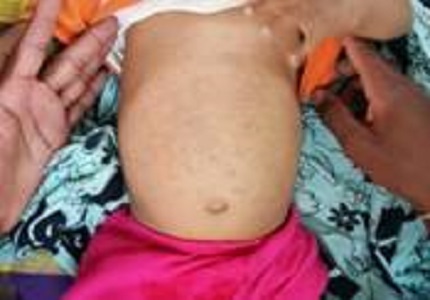
Extensive Mongolian spots: a clinical sign merits special attention for gm1-Gangliosidosis
Abstract
GM1-gangliosidosis (GM1) is one of the metabolic storage diseases, of which a differential diagnosis requires an array of biochemical assays to determine the enzyme deficiency. This approach is not only time-consuming and costly but also unavailable to most hospital laboratories. However, a presumptive diagnosis of GM1 may be made on the basis of diffuse ecchymosis, ectopic Mongolian spots and coarse facial feature, if present. A more definitive diagnosis of GM1 is then made on the demonstration of deficiency of GM1 beta-galactosidase in leukocytes, plasma or cultured skin fibroblasts. We postulate that dermal pigmentation may be recognized as an early sign of GM1 gangliosidosis.
Downloads
References
2. Carl Hayward, Hitesh C. Patel, Sanjay G. Manohar, Alexander R. Lyon. Gene therapy for GM1 gangliosidosis: challenges of translational medicine. Ann Transl Med. 2015 May; 3(Suppl 1): S28. [PubMed]
3. Shield JP, Stone J, Steward CG. Bone marrow transplantation correcting beta-galactosidase activity does not influence neurological outcome in juvenile GM1-gangliosidosis. J Inherit Metab Dis. 2005; 28(5):797-8.
4. Kasperzyk JL, El-Abbadi MM, Hauser EC, et al. N-butyldeoxygalactonojirimycin reduces neonatal brain ganglioside content in a mouse model of GM1 gangliosidosis. J Neurochem. 2004 May; 89(3):645-53. [PubMed]
5. Kasperzyk JL, d'Azzo A, Platt FM, et al. Substrate reduction reduces gangliosides in postnatal cerebrumbrainstem and cerebellum in GM1 gangliosidosis mice. J Lipid Res. 2005 Apr; 46(4):744-51. [PubMed]
6. Matsuda J, Suzuki O, Oshima A, et al. Chemical chaperone therapy for brain pathology in GM1-gangliosidosis. Proc Natl Acad Sci U S A. 2003 Dec;100(26):15912-7. [PubMed]
7. Suzuki Y, Ichinomiya S, Kurosawa M, et al. Chemical chaperone therapy: clinical effect in murine GM1- gangliosidosis. Ann Neurol. 2007 Dec; 62(6):671-5. [PubMed]
8. Landing BH, Silverman FN, Craig GM, Jacoby MD, Lahey ME, Chadwick DL. Familial neurovisceral lipidosis. Am J Dis Child. 1964 Nov;108:503–22. [PubMed]
9. Hooft C, Senesael L, Delbeke MJ, Kint J, Dacremont G. The Gm1 gangliosidosis (Landing disease). Eur Neurol. 1969; 2(4):225–41. [PubMed]
10. Ginsburg CM, Long CG. GM1 gangliosidosis type 1 in twins. J Med Genet. 1977 Apr;14(2):132–4. [PubMed]
11. Beratis N, Varvarigou-Frimas A, Beratis S, Sklower S. Angiokeratoma corporis diffusum in GM1 gangliosidosis, type 1. Clin Genet. 1989 Jul; 36(1):59–64. [PubMed]
12. Weissbluth M, Esterly NB, Caro WA. Report of an infant with GM1 gangliosidosis type I and extensive and unusual mongolian spots. Br J Dermatol. 1981 Feb;104(2):195–200. [PubMed]
13. Selsor LC, Lesher JL., Jr Hyperpigmented macules and patches in a patient with GM1 type 1 gangliosidosis. J Am Acad Dermatol. 1989 May;20(5 Pt 2):878–82. [PubMed]
14. Beattie RM, Harvey D. Extensive and unusual Mongolian blue spots in a child with GM1 gangliosidosis type one. J R Soc Med. 1992 Sep; 85(9):574–5. [PubMed]
15. Tang TT, Esterly NB, Lubinsky MS, Oechler HW, Harb JM, Franciosi RA. GM1-gangliosidosis type 1 involving the cutaneous vascular endothelial cells in a black infant with multiple ectopic mongolian spots. Acta Derm Venereol. 1993 Dec; 73(6):412–5. [PubMed]
16. Silengo M, Battistoni G, Spada M. Is there a relationship between extensive Mongolian spots and inborn error of metabolism? Am J Med genet. 1999 Nov; 87(3):276–7. [PubMed]
17. Ashrafi MR, Shabanian R, Mohammadi M, Kavusi S. Extensive Mongolian spots: a clinical sign merits special attention. Pediatr Neurol. 2006 Feb; 34(2):143–5. [PubMed]
18. Hanson M, Lupski JR, Hicks J, Metry D. Association of dermal melanocytosis with Lysosomal storage disease. Arch Dermatol. 2003 July; 139(7):916–20. [PubMed]
19. Ochiai T, Ito K, Okada T, Chin M, Shichino H, Mugishima H. Significance of extensive Mongolian spots in Hunter's syndrome. Br J Dermatol. 2003 June; 148(6):1173–8. [PubMed]
Copyright (c) 2016 Author (s). Published by Siddharth Health Research and Social Welfare Society

This work is licensed under a Creative Commons Attribution 4.0 International License.

 OAI - Open Archives Initiative