
Neuroimaging in Japanese encephalitis and their correlation with clinical profile in pediatric patients
Abstract
Introduction: Japanese encephalitis is a major public health problem in Indian subcontinent. Regardless of all advances in prompt diagnosis of JE, it may be difficult to differentiate JE from other viral encephalitis.
Aim: This study was done to know the topographic patterns of CT and MRI abnormalities in JE encephalitis.
Methodology: This retrospective observational study was done in children 1-15 years of age who suffered from JE encephalitis diagnosed by MAC-ELISA and in whom CT/MRI was done. Total 25 patients were enrolled. There CT/MRI findings were analysed and correlated with clinical features.
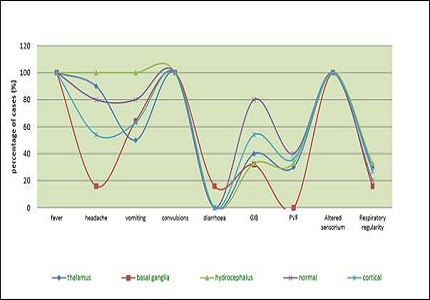
Results: The finding were principally seen in thalamic (40%; n=10) and basal ganglia (24%; n=6) in the form of hypo densities. Similar forms of lesions were also found in cortical region, Frontal=2 patients, parietal = 6 patients, temporal = 7 patients and occipital = 1 patient. MRI was done in eleven patients. Our MRI findings were also in correlation with CT findings with most common being thalamic (n=10) and basal ganglia (n=5). Temporal (n=4), parietal (n=3) and occipital (n=2) lobe changes.
Conclusions: The imaging findings on CT and MR imaging evidence the pathologic changes. Majority of lesions on CT/MRI were in thalamus and basal ganglia, but in some cases cortical regions were also involved. Temporal involvement, which was previously a reflection of Herpes encephalitis on CT/MRI, can also be seen in JE encephalitis.
Downloads
References
2. Diagana M, Preux PM, Dumas M. Japanese encephalitis revisited. J Neurol Sci. 2007 Nov 15;262(1-2):165-70. Epub 2007 Jul 23. [PubMed]
3. Yamanaka A, Mulyatno KC, Susilowati H, Hendrianto E, Utsumi T, Amin M, Lusida MI, Soegijanto S, Konishi E. Prevalence of antibodies to Japanese encephalitis virus among pigs in Bali and East Java, Indonesia, 2008. Jpn J Infect Dis. 2010 Jan;63(1):58-60. [PubMed]
4. van den Hurk AF, Ritchie SA, Mackenzie JS. Ecology and geographical expansion of Japanese encephalitis virus. Annu Rev Entomol. 2009;54:17-35. doi: 10.1146/annurev.ento.54.110807.090510. [PubMed]
5. Campbell GL, Hills SL, Fischer M, Jacobson JA, Hoke CH, Hombach JM, Marfin AA, Solomon T, Tsai TF, Tsu VD, Ginsburg AS. Estimated global incidence of Japanese encephalitis: a systematic review. Bull World Health Organ. 2011 Oct 1;89(10):766-74, 774A-774E. doi: 10.2471/BLT.10.085233. Epub 2011 Aug 3.
6. A Mathur, K L Arora, S Rawat, and U C Chaturvedi. “Persistence, latency and reactivation of Japanese encephalitis virus infection in mice,” Journal of General Virology. 1986;67:381–385. [PubMed]
7. Saxena SK, Singh M, Pathak AK, Mathur A. Reply to 'Encephalitis outbreak finds Indian officials unprepared'. Nat Med. 2006 Mar;12(3):269-70.
8. Kalita J, Misra UK. Comparison of CT scan and MRI findings in the diagnosis of Japanese encephalitis. J Neurol Sci. 2000 Mar 1;174(1):3-8. [PubMed]
9. Kalita J, Misra UK, Pandey S, Dhole TN. A comparison of clinical and radiological findings in adults and children with Japanese encephalitis. Arch Neurol. 2003 Dec;60(12):1760-4. [PubMed]
10. Shoji H, Kida H, Hino H, Matsuura S, Kojima K, Abe T, Utsunomiya H, Okada Y, Nakamura Y, Okudera T. Magnetic resonance imaging findings in Japanese encephalitis. White matter lesions. J Neuroimaging. 1994 Oct;4(4):206-11.
11. Kalita J, Misra UK. Comparison of CT scan and MRI findings in the diagnosis of Japanese encephalitis. J Neurol Sci. 2000 Mar 1;174(1):3-8. [PubMed]
12. Handique SK, Das RR, Barman K, Medhi N, Saharia B, Saikia P, Ahmed SA. Temporal lobe involvement in Japanese encephalitis: problems in differential diagnosis. AJNR Am J Neuroradiol. 2006 May;27(5):1027-31. [PubMed]
13. Gourie-Devi M, Ravi V, Shankar SK. Japanese encephalitis. An overview.In: Clifford Rose, ed. Recent advances in tropical neurology. Amsterdam: Elsevier Science B.V;1995:217–235. [PubMed]
14. Mohr JP, Steinke W, Timsit SG, Sacco RL, Tatemichi TK. The anterior choroidal artery does not supply the corona radiata and lateral ventricular wall. Stroke. 1991 Dec;22(12):1502-7. [PubMed]
15. Osborne AG, Davis WL, Jacobs J. Normal vascular anatomy. In: Osborne AG, ed. Diagnostic neuroradiology. St. Louis, Mo: Mosby-Year Book, Inc.; 1994:117–53. [PubMed]
16. Bressman SB, de Leon D, Raymond D, Greene PE, Brin MF, Fahn S, Ozelius LJ, Breakefield XO, Kramer PL, Risch NJ. Secondary dystonia and the DYTI gene. Neurology. 1997 Jun;48(6):1571-7. [PubMed]
17. Ceballos-Baumann AO, Passingham RE, Marsden CD, Brooks DJ. Motor reorganization in acquired hemidystonia. Ann Neurol. 1995 Jun;37(6):746-57.
18. Wooten GF, Lopes MB, Harris WO, Reagan TJ, Vandenberg SR. Pallidoluysian atrophy: dystonia and basal ganglia functional anatomy. Neurology. 1993 Sep;43(9):1764-8.
Copyright (c) 2016 Author (s). Published by Siddharth Health Research and Social Welfare Society

This work is licensed under a Creative Commons Attribution 4.0 International License.

 OAI - Open Archives Initiative