Role of ultra sonography (chest and abdomen) in diagnosis and early prediction of severity of dengue fever
Abstract
Introduction: The diagnosis of Dengue fever is often delayed owing to time taken for availability of serology test results. Ultrasonography (USG) is a cheap, rapid and widely available non-invasive imaging method. Aim of the study was to access the role of ultrasonographic features of thorax and abdomen in diagnosis and early prediction of severity of dengue fever.
Material and Method: It was aobservational descriptive study, conducted during the period of September 2017 to august 2018 at department of Paediatrics, J. K. Lon Mother and Child Hospital, Government Medical College, Kota. Out of 122 suspected dengue fever cases of age group 2 month to 18 years, 84 children were seropositive for dengue fever, were sent for Ultrasound scan of the abdomen and thorax.
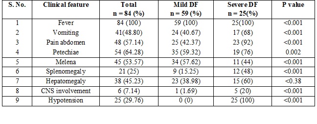
Result: 59(70.23%) cases were in mild dengue group and 25(29.76%) cases were in severe dengue group. All cases had fever. About 41(57.14%) cases had vomiting, 48(57.14%) had pain abdomen, 54 (64.28%) had Petechiae, 45 (53.57%) had melena. 38 (45.23%) had hepatomegaly, 21 (25%) had splenomegaly and 25 (29.76%) had hypotension. Gall bladder wall edema, ascites, pleural effusion, hepatomegaly, splenomegaly and perinephric edema were present in 67 (79.76%), 52 (61.9%), 43 (51.19%), 51 (60.71%), 27 (32.14%) and 14 (16.66%) in all dengue fever group while 25 (100%), 25 (100%), 21 (84%), 15 (60%), 10 (40%), and 11 (44%) in severe dengue group respectively. All sonographic features had more significant association with severe dengue group (p <0.001) except hepatomegaly. All sonographic features had significant correlation (P value < 0.001) with severe thrombocytopenia except hepatomegaly.
Conclusion: Ultrasonography is a simple and valuable tool in diagnosing and predicting severity of dengue fever.
Downloads
References
2. World Health Organization. Health Situation in South-East Asia Region 2001–2007. New Delhi, India: World Health Organization; 2008.
3. World Health Organization. Monograph on Dengue / Dengue Haemorrhagic fever. Regional Publication, World Health Organization. 1983; SEARO. 22
4. Rigau-Pérez JG, Clark GG, Gubler DJ, et al. Dengue and dengue haemorrhagic fever. Lancet. 1998 Sep 19;352(9132):971-7.[pubmed]
5. Handbook for clinical management of dengue fever. WHO; 2012.
6. Bandyopadhyay S, Lum LC, Kroeger A. et al. Classifying dengue: a review of the difficulties in using the WHO case classification for dengue haemorrhagic fever. Trop Med Int Health. 2006 Aug;11(8):1238-55. DOI:10.1111/j.1365-3156.2006.01678.x.[pubmed]
7. Venkata Sai PM, Dev B, Krishnan R. et al. Role of ultrasound in dengue fever. Br J Radiol. 2005 May;78(929):416-8.[pubmed]
8. Thulkar S, Sharma S, Srivastava DN, et al. Sonographic findings in grade III dengue hemorrhagic fever in adults. J Clin Ultrasound. 2000 Jan;28(1):34-7.[pubmed]
9. NELSON ER. Hemorrhagic fever in children in Thailand. Report of 69 cases. J Pediatr. 1960 Jan;56:101-8.[pubmed]
10. WHO (1997). "Chapter 2: clinical diagnosis"(PDF). Dengue haemorrhagic fever: diagnosis, treatment, prevention and control (2nd ed.). Geneva: World Health Organization. pp. 12–23.
11. Ukey P, Bondade S, Paunipagar P, et al.Study of seroprevalence of dengue Fever in central India. Indian J Community Med. 2010 Oct;35(4):517-9. doi: 10.4103/0970-0218.74366.[pubmed]
12. Srivastava VK, Suri S, Bhasin A, et al. An epidemic of dengue haemorrhagic fever and dengue shock syndrome in Delhi: a clinical study. Ann Trop Paediatr. 1990;10(4):329-34.[pubmed]
13. Bethell DB, Gamble J, Loc PP, Dung NM, Chau TTH, Loan HT, et al. Non-invasive measurement of microvascular leakage in patients with dengue hemorrhagic fever. Clin Infect Dis 2001 Jan 15;32(2): 243-53. Epub 2001 Jan 15.
14. Aggarwal A, Chandra J, Aneja S, et al. An epidemic of dengue hemorrhagic fever and dengue shock syndrome in children in Delhi. Indian Pediatr. 1998 Aug;35(8):727-32.[pubmed]
15. Pushpa V, Venkatadesikalu M, Mohan S, et al. An epidemic of dengue haemorrhagic fever/dengue shock syndrome in tropical India. Ann Trop Paediatr. 1998 Dec;18(4):289-93.[pubmed]
16. Sachar S, Goyal S, Sacha S. Role of Ultrasonography (“Honeycomb Sign”) in Early Detection of Dengue Hemorrhagic Fever. Arch Clin Exp Surg. 2013;2(1):38-42
17. Williandry M, Laraswati B. Profile of Chest and Abdomen Ultrasound on Patients with Dengue Virus Infection. Folia Medica Indonesiana 2013 December;49(4):237-43.
18. Santhosh VR, Patil PG, Srinath MG, et al. Sonography in the diagnosis and assessment of dengue Fever. J Clin Imaging Sci. 2014 Mar 21;4:14. doi: 10.4103/2156-7514.129260. eCollection 2014. [pubmed]
Copyright (c) 2019 Author (s). Published by Siddharth Health Research and Social Welfare Society

This work is licensed under a Creative Commons Attribution 4.0 International License.

 OAI - Open Archives Initiative