
Role of cranial ultrasound for diagnosis of intracranial abnormalities in newborns
Abstract
Aim: To evaluate the role of cranial ultrasound in neonates and infants in diagnosis of various intracranial abnormalities.
Method: A real time cranial ultrasound was performed on 98 neonates and 33 infants of 1 month to 1 year of age group including the all preterms and symptomatic term neonates and infants during the study period.
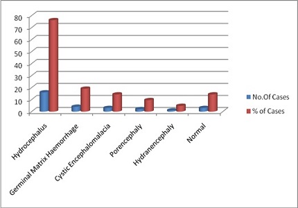
Result: Sonography results in symptomatic premature neonates showed Germinal Matrix haemorrhage in 67.56%, hydrocephalus in 37.83% and peri-ventricular leukomalacia in 24.31%, while in asymptomatic premature newborn, sonography showed germinal matrix haemorrhage/hydrocephalus in 27.5% and normal scan in rest of them. In symptomatic full term neonates, sonography showed hydrocephalus in 76.19%, germinal matrix hemorrhage in 19.04%, cystic encephalomalacia in 14.28%, porencephaly in 9.52%, hydrancephaly in 4.76% and normal scan in 14.28%. Sonographic scan in symptomatic infants of more than 1 month of age showed – echogenic sulci in 39.39% cases, hydrocephalus in 33.33%, cerebritis in 15.15%, cerebral abscess in 6.06%, mass lesions in 3.03% and normal scan in 24.24% cases.
Conclusion: Because of being safe, nonionising and easily available, cranial ultrasound can be used as a primary imaging modality to detect intracerebral lesions in neonates and infants.
Downloads
References
2. Lawn JE, Cousens S, Zupan J; Lancet Neonatal Survival Steering Team. 4 million neonatal deaths: when? Where? Why? Lancet. 2005 Mar 5-11;365(9462):891-900. [PubMed]
3. Lawn JE, Cousens SN, Wilczynska K. Estimating the causes of four million neonatal deaths in the year 2000 : statistical annex. In : The world health report 2005. Geneva: WHO; 2005.
4. ICMR Young Infant Study Group. Age profile of neonatal deaths. Indian Pediatr. 2008 Dec;45(12):991-4. [PubMed]
5. T. Bang, V. K. Paul, H. M. Reddy, and S. B. Baitule, Why do neonates die in rural Gadchiroli, India? (Part I): primary causes of death assigned by neonatologist based on prospectively observed records, Journal of Perinatology , vol . 25 , supplement 1 , pp. S29–S34, 2005. View at Publisher View at Google Scholar.
6. Adams C, Johnston WP, Nevin NC. Family study of congenital hydrocephalus. Dev Med Child Neurol. 1982 Aug;24(4):493-8. [PubMed]
7. Armstrong DL, Sauls CD, Goddard-Finegold J. Neuropathologic findings in short-term survivors of intraventricular hemorrhage. Am J Dis Child. 1987 Jun;141(6):617-21. [PubMed]
8. Ambrosino MM, Hernanz-Schulman M, Genieser NB, Wisoff J, Epstein F. Brain tumors in infants less than a year of age. Pediatr Radiol. 1988;19(1):6-8. [PubMed]
9. Dewbury KC, Aluwihare AP. The anterior fontanelle as an ultrasound window for study of the brain: a preliminary report. Br J Radiol. 1980 Feb;53(626):81-4. [PubMed]
10. Sonography of the neonatal brain” Journal of Diagnostic Medical Sonography Volume 25, Issue 6, November2009,Pages331-348 DOI: 10.1177/8756479309347801.
11. Levene MI. Measurement of the growth of the lateral ventricles in preterm infants with real-time ultrasound. Arch Dis Child. 1981 Dec;56(12):900-4. [PubMed]
12. Rumack CM, Drose JA. Neonatal and infant brain imaging. In: Rumack CM, Wilson SR, Charboneau JW (eds). Diagnostic Ultrasound. Vol 2. 4th ed. Philadelphia, PA: Elsevier; 2011:1558–1636. [PubMed]
13. Siegel MJ. Brain. In: Siegel MJ (ed). Pediatric Sonography. 4th ed. Philadelphia, PA: Lippincott Williams & Wilkins; 2011:43–117. 5. Slovis TL, Bulas DI, Nelson MD. Neonatal brain imaging. [PubMed]
14. Slovis TL, Coley BD, Bulas DI, et al (eds). Caffey’s Pediatric Diagnostic Imaging. Vol 1. Philadelphia, PA: Elsevier; 2008:398–429.
15. Kristin Fickenscher, Zachary Bailey, Megan Saettele, Amy Dahl and Lisa Lowe (2012). Pediatric Cranial Ultrasound: Techniques, Variants and Pitfalls, Neuroimaging - Methods, Prof. Peter Bright (Ed.), ISBN:978-953-51-0097-3.
16. BANKER BQ, LARROCHE JC. Periventricular leukomalacia of infancy. A form of neonatal anoxic encephalopathy. Arch Neurol. 1962 Nov;7:386-410. [PubMed]
17. Barkovich AJ, Kjos BO, Norman D, Edwards MS. Revised classification of posterior fossa cysts and cystlike malformations based on the results of multiplanar MR imaging. AJR Am J Roentgenol. 1989 Dec;153(6):1289-300. [PubMed]
18. Barson AJ. Spina bifida: the significance of the level and extent of the defect to the morphogenesis. Dev Med Child Neurol. 1970 Apr;12(2):129-44. [PubMed]
19. Mercuri E, Dubowitz L, Brown SP, Cowan F: incidence of cranial ultrasound abnormalities in neonates and association with post natal factors and neurological status. Arch Dis Child Fetal Neonatal Ed 1998; 79(3):F185-9.
20. Sims ME, Turkel SB, Halterman G, Paul RH. Brain injury and intrauterine death. Am J Obstet Gynecol. 1985 Mar 15;151(6):721-3. [PubMed]
21. Perry RN, Bowman ED, Murton LJ, Roy RN, de Crespigny L. Cranial ultrasound screening of preterm and term neonates. Aust Paediatr J. 1987 Feb;23(1):31-3. [PubMed]
22. Ment LR, Duncan CC, Scott DT, Ehrenkranz RA. Posthemorrhagic hydrocephalus. Low incidence in very low birth weight neonates with intraventricular hemorrhage. J Neurosurg. 1984 Feb;60(2):343-7. [PubMed]
23. Edwards MK, Brown DL, Muller J, Grossman CB, Chua GT. Cribside neurosonography: real-time sonography for intracranial investigation of the neonate. AJR Am J Roentgenol. 1981 Feb;136(2):271-5. [PubMed]
Copyright (c) 2016 Author (s). Published by Siddharth Health Research and Social Welfare Society

This work is licensed under a Creative Commons Attribution 4.0 International License.

 OAI - Open Archives Initiative