Study of transcranial colour Doppler in the measurement of cerebral edema in birth asphyxia
Abstract
Background: Perinatal asphyxia is an insult to fetus or newborn due to lack of oxygen /perfusion to various organs. Perinatal asphyxia occurs in 1 to 3 per 1000 live births and is related to gestational age and birth weight. Worldwide 4-9 million suffer from birth asphyxia. It occur in 9% of infants of <36 wks of gestation and 0.5% in > 36 wks. Hypoxic ischemic brain injury is the most important consequence of perinatal asphyxia.
Aim of study: To detect the measurement of cerebral blood flow in full term infants with birth asphyxia, by use of transcranial colour Doppler to measure RI (Resistive Index).
Setting: Department of Paediatrics at MY hospital in collaboration with department of radiodiagnosis over a period of 1 yr from November 2013- October 14.
Design: Observational study.
Method: It included 40 consecutive cases of birth asphyxia who suffered from HIE. Measurement of cerebral blood flow velocity was done by the use of colour doppler ultrasound.
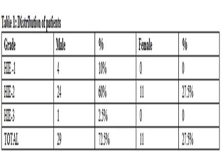
Result: Resistive index values were lower in measured vessels in asphyxiated infant as compared by blood flow velocity, resistive index of ACA and MCA. It is observed in our study that maximum number of patients in asphyxiated group belongs to grade II HIE 87.5% (35), while in stage III there were 2.5% (1) and in stage I only 4 (10%) patients.
Conclusion: Colour Doppler USG would be of practical importance in evaluating the cerebral blood flow velocity in neonate with HIE. A skillful detection of decrease in cerebral blood flow of neonate in postnatal first 12 hrs & treatment being based on such detection would contribute to a good prognosis.
Downloads
References
2 . Vannucci RC Hypoxic ischemic encephalopathy –American Journal of perinatology DOI: 10.1055/s-2000-9293. [PubMed]
3. Judith H Meek, Clare E Elwell, David C McCormick, A David Edwards, Janice P Townsend, Ann L Stewart, John S Way ADC. fetal and neonatal edition- abnormal cerebral hemodynamics in perinatally asphyxiated infants and their outcome. Arch Dis Child Fetal Neonatal Ed. 1999 Sep; 81(2): F110–F115.
4. van Handel M, Swaab H, de Vries LS, Jongmans MJ. Long-term cognitive and behavioral consequences of neonatal encephalopathy following perinatal asphyxia: a review. Eur J Pediatr. 2007 Jul;166(7):645-54. Epub 2007 Apr 11.
5. Elstad M, Whitelaw A, Thoresen M. Cerebral Resistance Index is less predictive in hypothermic encephalopathic newborns. Acta Paediatr. 2011 Oct;100(10):1344-9. doi: 10.1111/j.1651-2227.2011.02327.x. Epub 2011 May 18. [PubMed]
6. Judith H Meek, Clare E Elwell, David C McCormick, A David Edwards, Janice P Townsend, Ann L Stewart, John S Way; abnormal cerebral hemodynamics in perinatally asphyxiated infants and their outcome. Arch Dis Child Fetal Neonatal Ed. 1999 Sep; 81(2): F110–F115.
7. Badawi N, Kurinczuk JJ, Keogh JM, Alessandri LM, O'Sullivan F, Burton PR, Pemberton PJ, Stanley FJ. Intrapartum risk factors for newborn encephalopathy: the Western Australian case-control study. BMJ. 1998 Dec 5;317(7172):1554.
8. van Bel F, van de Bor M, Stijnen T, Baan J, Ruys JH. Cerebral blood flow velocity pattern in healthy and asphyxiated newborns: a controlled study. Eur J Pediatr. 1987 Sep;146(5):461-7. [PubMed]

Copyright (c) 2016 Author (s). Published by Siddharth Health Research and Social Welfare Society

This work is licensed under a Creative Commons Attribution 4.0 International License.


 OAI - Open Archives Initiative


