
Sonographic features of neonatal intestinal malrotation and volvulus
Abstract
Background: Normally third portion of duodenum is always retromesenteric-retroperitoneal. Demonstration of intramesentric position of D3 on ultrasound can help in diagnosis of malrotation.
Objective: The aim of this study was to evaluate the feasibility of Ultrasound in demonstrating the retroperitoneal D3 for early and effective diagnosis of malrotation.
Methods: A 5-year prospective observational study was done between April 2011 to March 2016 in the neonatal ICU of Vydehi institute of medical sciences. 122 neonates presented with features of vomiting and abdominal distension. After ruling out all medical and surgical causes (other) possibility of malrotation was suspected in 41 babies were included in the study. Plain abdominal radiography, sonography and upper gasto intestinal barium examinations were performed in all the 41 suspected cases.
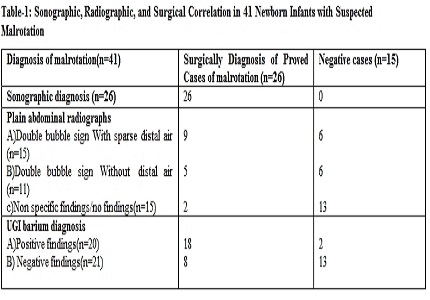
Results: All the 41 cases were evaluated and later surgical intervention was done. 26 out of 41 cases had ultrasonographic features of malrotation, which were subsequently proved by surgery. Inversions of Superior mesenteric artery and Superior mesenteric vein were found in all the 26 cases that were surgically proved of malrotation. Other features like thickened duodenal wall greater than 2mm in 16 cases, intraperitoneal location of third part of duodenum in 13 cases, distal dilatation of the SMV in 11 cases, duodenal dilatation with tapering configuration in 10 cases and ascites in 14 cases were found.
Conclusion: Ultrasonography provides good diagnostic results in neonatal intestinal malrotation. Specific Sonographic features relating to volvulus should be evaluated as potential indicators of the need for an emergent operation.
Downloads
References
2. Simpson AJ, Leonidas JC, Krasna IH, et at: Roentgen diagnosis of midgut malrotation: Value of upper gastrointestinal radiographic study. J Pediatr Surg.1972 Jul; 243(7):154-9.
3. Ford EG, Senac MO Jr, Srikanth MS, Weitzman JJ. Malrotation of the intestine in children. Ann Surg. 1992 Feb;215(2):172-8. [PubMed]
4. Loyer E, Dunne Eggli K.Sonographic evaluation of superior mesenteric vascular relationship in malrotation. Pediatr Radiol.1989 Mar;19(3):173-5.
5. R Yanez, L Spitz.Intestinal malrotation presenting outside the neonatal period.Archives of disease in childhood.1986 Jul;71(7):682-685.
6. Long FR, Kramer SS, Markowitz RI .Intestinal malrotation in children: tutorial on radiographic diagnosis in difficult cases. Radiology. 1996 Mar;198(3):775-80. Doi:101148/radiology.198.3.8628870.
7. Mori H, Hayashi K, Futagawa S, Uetani M, Yanagi T, Kurosaki N. Vascular compromise in chronic volvulus with midgut malrotation. Pediatr Radiol. 1987;17(4):277-81.
8. Weinberger E, Winters WD, Liddell RM, Rosenbaum DM, Krauter D. Sonographic diagnosis of intestinal malrotation in infants: importance of the relative positions of the superior mesenteric vein and artery. AJR Am J Roentgenol. 1992 Oct;159(4):825-8.
9. Patino MO, Munden MM. Utility of the sonographic whirlpool sign in diagnosing midgut volvulus in patients with atypical clinical presentations. J Ultrasound Med. 2004 Mar;23(3):397-401.
10. Berdon WE. The diagnosis of malrotation and volvulus in the older child and adult: a trap for radiologists. Pediatr Radiol. 1995;25(2):101-3. [PubMed]
Copyright (c) 2017 Author (s). Published by Siddharth Health Research and Social Welfare Society

This work is licensed under a Creative Commons Attribution 4.0 International License.

 OAI - Open Archives Initiative